-
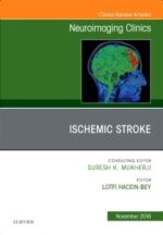
Neurografía por RM Plexos y nervios periféricos – Cejas
$385,000La resonancia magnética es una técnica de imagen que pareciera no haber alcanzado aún su límite y la neurografía por RM es uno de sus más importantes avances en los últimos años. Se la considera una herramienta diagnóstica complementaria y no invasiva para evaluar la patología de nervios y plexos, que permite la visualización directa de las estructuras nerviosas brindando una mayor exactitud diagnóstica respecto a otros métodos de estudio.
El libro es un tratado completo sobre neurografía por RM, ilustrado bellamente con más de 500 imágenes. Presenta los temas de forma clara, concisa y fácil de entender. Los primeros capítulos están organizados por nervios específicos y desarrollan la anatomía básica y su fisiología, las evaluaciones clínicas, los protocolos de imagen y la patología por regiones anatómicas. Los capítulos que siguen se focalizan en los nervios craneales y en el desarrollo exhaustivo de la patología tumoral en nervios y plexos, que incluye un apartado sobre neurofibromatosis. Para el cierre, se destina un capítulo al tratamiento quirúrgico de muchas de las condiciones discutidas.
$453,000 -
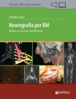
Ecografía Transvaginal en la Evaluación de los Tumores de Ovario (incluye versión digital)
$117,000La ecografía transvaginal es una de las pruebas de diagnóstico por imagen más importantes para el ginecólogo a la hora de discriminar las lesiones benignas de las malignas que surgen en los anejos uterinos (ovario y trompas de Falopio). Aunque se trata de una técnica de imagen utilizada en este escenario clínico desde hace más de tres décadas su evolución permite, en la actualidad, una evaluación estandarizada, el desarrollo de modelos matemáticos predictivos y la incorporación de avances tecnológicos, como la ecografía 3D/4D.
$138,000 -
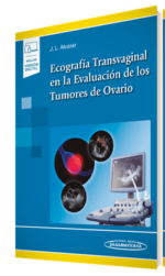
Ecografía 4ª Ed. 2 Volúmenes – Rumack
$841,000Ecografía de Rumack es el tratado de ultrasonografía más completo y actualizado del mercado.
Está dirigido a radiólogos, residentes en formación y todos los especialistas interesados en conocer las aplicaciones de la ecografía diagnóstica.
Esta 4ª edición cuenta con más de 5.000 nuevas ilustraciones, pero no se trata solo de una colección de nuevas imágenes de altísima calidad, comentadas por los más reconocidos especialistas en ecografía, el Rumack antes de facilitar el diagnóstico de una determinada patología, aborda la anatomía normal, las posibles variantes anatómicas e incluso la histología que marca diferentes patrones ecográficos. Después analiza a fondo las diferentes imágenes de cada caso, aportando diagnósticos diferenciales, errores frecuentes, recomendaciones de pruebas alternativas y técnicas intervencionistas.
En todos los capítulos se incluyen figuras con varias exploraciones sumamente útiles o composiciones de características fundamentales. Todas estas imágenes reflejan el espectro de alteraciones ecográficas que pueden aparecer en una enfermedad, en lugar de únicamente la manifestación más frecuente.
La revisión y puesta al día de esta obra son realmente un acontecimiento, que han hecho del Rumack el libro más deseado y utilizado de ecografía en el mundo.
$990,000 -
Learning Interventional Radiology
$240,000Now designated as a primary medical specialty, the field of interventional radiology has contributed many ground-breaking procedures, including angioplasty, catheter-delivered stents, aneurysm coiling, and minimally-invasive cancer treatment. This first-of-its-kind review text offers an authoritative, easy-to-use introduction to the field, highlighting procedures, instruments, techniques, modalities, and more. Using an image-filled, practical format it covers exactly what you need to know for a solid foundation in this fast-growing field.
-
Brain Embryology and the Cause of Congenital Malformations, An Issue of Neuroimaging Clinics
$360,000This issue of Neuroimaging Clinics of North America focuses on Brain Embryology and the Cause of Congenital Malformations and is edited by Drs. Thierry A.G.M. Huisman and Avner Meoded. Articles will include: Neuroimaging of normal brain development; Ultrasound and MRI of the normal fetal brain; Spinal dysraphia, Chiari 2 malformation, unified theory and advances in fetoscopic repair; Posterior fossa malformations; Synopsis of brain embryology; Cerebral dysplasia and overgrowth syndromes; Disorders of ventral induction/spectrum of holoprosencephaly; Classification and neurogenetics of intracranial vascular anomalies; DTI of brain malformations: Exploring the internal architecture; Connectomics in brain malformations: How is the malformed brain wired?; Commissural anomalies; and more!
-
Top 3 Differentials in Nuclear Medicine A Case Review
$288,000The highest-yield, most complete nuclear radiology exam prep and learning tool available today!
Top 3 Differentials in Nuclear Medicine: A Case Review by renowned nuclear radiologist Ely A. Wolin and esteemed contributors is one in a series of radiology case books mirroring the format of the highly acclaimed O’Brien classic, Top 3 Differentials in Radiology: A Case Review. The book is organized into 12 parts, with initial parts covering neuro, thyroid and parathyroid, cardiac, lung, hepatobiliary, gastrointestinal, genitourinary, and bone imaging. Latter parts focus on imaging of various inflammatory processes, infections, and neoplasms. The final part covers the important topic of quality control, which is essential for both American Board of Radiology (ABR) exam review and clinical practice.
Each case is formatted as a two-page unit. The left page features clinical images, succinctly captioned findings, and pertinent clinical history. The right page includes the key imaging gamut, differential diagnoses rank-ordered by the «Top 3,» additional diagnostic considerations, and clinical pearls.
-
PET Correlación con TC y RM – Bennet
$571,000PET: Correlación con TC y RM es la primera guía completa centrada en el diagnóstico sobre el uso efectivo de la emergente tecnología PET. Es también un recurso integral adaptado para apoyar la toma de decisiones en el lugar de atención. Este texto accesible y de referencia contiene todo lo que se necesita saber acerca del papel fundamental de la PET en diferentes áreas: oncología; cardiología; infección e inflamación; patología vascular, mamaria, neurológica, musculoesquelética, gastrointestinal, neuroendocrina y muchas otras. Esta edición incluye: – Más de 1600 imágenes de alta calidad con epígrafes y anotaciones para guiar la interpretación mediante ilustraciones, imágenes PET y correlación con TC y RM. – Listas de diagnósticos para una referencia rápida y fácil. – Capítulos sobre interpretación por parte de especialistas, artefactos y dificultades frecuentes. – Un amplio espectro de información esencial, como diagnósticos oncológicos mediante PET con tablas de estadificación y recomendaciones para el informe; indicaciones cardíacas para PET (pruebas de esfuerzo, viabilidad miocárdica, sarcoidosis, etc.); indicaciones de la PET en SNC (demencia, epilepsia) y oncología, entre otras; y casos pedagógicos ilustrados que incluyen la correlación mediante TC y RM. – Física e instrumentación de la PET. Presentación de los radiotrazadores clínicos y emergentes en formato de tabla. Ideal para médicos que atienden pacientes con cáncer -radiólogos, médicos nucleares, radioterapeutas, oncólogos, cirujanos y profesionales en formación en medicina nuclear y oncología- así como aquellos profesionales que interpretan PET por una amplia variedad de indicaciones. Esta versión impresa incluye el acceso gratuito a la versión electrónica
$672,000 -
Radiología Esencial 2Ed. (incluye eBook) 2 TOMOS
$1,016,000La publicación de esta 2ª edición de Radiología Esencial es consecuencia, por un lado, del enorme éxito de la 1ª edición de la obra y, por otro, de la necesidad de renovar sus contenidos dados los avances científicos que se han producido en el campo de la Radiología en los últimos años.
La Radiología es una especialidad médica en constante crecimiento que se ha convertido en la técnica paraclínica más relevante para establecer diagnósticos precisos y precoces en un elevadísimo número de procesos. Además, juega un papel esencial en la monitorización de múltiples enfermedades con el objeto de analizar su evolución y respuesta terapéutica.
$1,195,000 -
Vascular and Interventional Radiology: A Core Review
$245,000Designed specifically for the Core Exam, Vascular and Interventional Radiology : A Core Review covers all key aspects of the field, mimicking the image-rich, multiple-choice format of the actual test. Ideal for residents preparing for the Core Examination, as well as practitioners taking recertification exams, this unique review follows the structure and content of what you’ll encounter on the test, effectively preparing you for Core Exam success!
- Contains 300 questions with answers, explanations, and references.
- Features high-quality angiographic, fluoroscopic, CT, MRI, and ultrasound images that demonstrate the broad range of clinical entities encountered in vascular and interventional radiology.
- Provides concise answers that include rationales, clinical pearls, and literature references, as well as high-yield tables embedded in the answers for additional review.
Enhance Your eBook Reading Experience:
- Read directly on your preferred device(s), such as computer, tablet, or smartphone.
- Easily convert to audiobook, powering your content with natural language text-to-speech.
-
Cranial Neuroimaging and Clinical Neuroanatomy
$869,000Thieme’s classic, indispensable guide to sectional imaging of the cranium
Now in a revised and expanded fourth edition, this exquisitely illustrated text/atlas by renowned experts, provides you with the cognitive tools to visualize and interpret CT and MR images of the cranium. In exacting detail, the normal structures of the brain, as seen in the three orthogonal planes (axial, sagittal, and coronal), are revealed with unparalleled accuracy, making the volume a highly useful aid in daily practice, for teaching, and to provide an anatomic baseline for research on the brain.
Beyond the clinical utility of the contents, the work is an aesthetic pleasure to behold, making learning and comprehension of complex material as simple and easy as possible.
-
Pediatric MR Imaging, An Issue of Magnetic Resonance Imaging Clinics of North America
$387,000This issue of MRI Clinics of North America focuses on Pediatric MR Imaging, and is edited by Dr. Edward Y. Lee. Articles will include: MRI Evaluation of Pediatric Neck Masses: Review and Update; MRI of Lungs and Airways in Children: Past and Present; Pediatric Mediastinal Masses: Role of MRI As a Problem-Solving Tool; Pediatric Cardiac MRI: Practical Preoperative Assessment; Hepatobiliary MRI in Children: Up-To-Date Imaging Techniques and Findings; Pediatric Renal Neoplasms: MRI-Based Practical Diagnostic Approach; MRI Evaluation of Inflammatory Bowel Disease in Children: Where Are We Now in 2018?; MRI Evaluation of Pediatric Genital Disorders: MR Technology Overview and Interpretation; Pediatric Sport-related Injuries: An Imaging Overview for Current and Future Daily Practice; MRI of Pediatric Musculoskeletal Tumors: Recent Advances and Clinical Applications; MRI Evaluation of Pediatric Lymphatics: Overview of Techniques and Imaging Findings; PET-MRI: Current Updates on Pediatric Applications; Tales from the Night: Emergency MRI in Pediatric Patients after Hours; and more!
-
Problem Solving in Chest Imaging
$733,000Optimize diagnostic accuracy with Problem Solving in Chest Imaging, a new volume in the Problem Solving in Radiology series. This concise title offers quick, authoritative guidance from experienced radiologists who focus on the problematic conditions you’re likely to see-and how to reach an accurate diagnosis in an efficient manner.
Key Features
-
- Addresses the practical aspects of chest imaging-perfect for practitioners, fellows, and senior level residents who may or may not specialize in chest radiology, but need to use and understand it.
- Helps you make optimal use of the latest imaging techniques and achieve confident diagnoses.
- Presents content by organ system and commonly encountered problems, with problem solving techniques integrated throughout.
-
- Features more than 1,500 high-quality images that provide a clear picture of what to look for when interpreting studies.
-
- Focuses on the core knowledge needed for successful results, covering anatomy, imaging techniques, imaging approach, entities by pathologic disease and anatomic region, and special situations. Key topics include Diffuse Lung Disease, Neoplasms of the Lung and Airways, Interstitial Lung Disease, Smoking-Related Lung Diseases, and Cardiovascular Disease.
- Shows how to avoid common problems that can lead to an incorrect diagnosis. Tables and boxes with tips, pitfalls, and other teaching points show you what to look for, while problem-solving advice helps you make sound clinical decisions.
- Expert Consult™ eBook version included with purchase. This enhanced eBook experience allows you to search all of the text, figures, and references from the book on a variety of devices.
-
-
Fundamentals of Body CT, 5th Edition – Webb
$230,000From recent advances in helical CT techniques to new developments in lung cancer screening to optimized CT techniques in musculoskeletal diagnosis, Fundamentals of Body CT, 5th Edition, covers the essential information you need to know to effectively perform and interpret CT scans. Step-by-step instructions for all current CT techniques help you quickly understand each procedure and review key steps. Comprehensive and easy to digest, this introduction to body CT is an essential resource for radiology residents, practicing radiologists, and medical students.
Key Features
-
- Features many new topics, discussions of additional diseases, and new, high-quality images from cover to cover, including updated descriptions and illustrations of normal anatomy and incidental findings.
- Allows you to quickly compare diagnoses with a survey of major CT findings for a variety of common diseases―with an emphasis on those findings that help to differentiate one condition from another.
- Reviews the spiral/helical CT protocols currently used for the diagnosis of chest, abdominal, and musculoskeletal abnormalities, including high-resolution CT, lung nodule assessment and lung cancer screening, CT pulmonary embolism diagnosis, CT enterography, CT enteroclysis, CT colonography, and optimizing CT techniques in musculoskeletal diagnosis.
- Brings you up to date with recent advances in chest CT, including the classification of adenocarcinoma, evaluation of lung nodules, lung cancer screening (including Lung-RADS) and staging, and the classification and diagnosis of interstitial lung diseases using high-resolution CT
- Covers new developments in abdominal CT such as the Liver Imaging Reporting and Data System (Li-RADS) for imaging and reporting small hepatocellular carcinoma, reviews of the Atlanta Classification of Acute Pancreatitis, and an improved description of CT findings of histologic subtypes of renal cell carcinoma.
- Includes new discussions of the diagnosis of musculoskeletal abnormalities detected on chest and abdominal CT scans obtained for non-musculoskeletal indications.
- Contains updated disease classifications, including those for pulmonary adenocarcinoma, diffuse lung diseases, and pancreatic lesions.
- Enhanced eBook version included with purchase. Your enhanced eBook allows you to access all of the text, figures, and references from the book on a variety of devices.
$459,000 -
-
Topics in Spine Imaging, An Issue of Radiologic Clinics of North America
$452,000This issue of Radiologic Clinics of North America focuses on The Spine, and is edited by Dr. Lubdha M. Shah. Articles will include: Pearls and Pitfalls in Spine Imaging; Traumatic Spinal Cord; Vascular Disorders of the Spine; Approach to (acute, subacute, chronic) Myelopathy; Acute Low Back Pain; Spinal Manifestations of Systemic Disease; Intraspinal Tumors; Spinal Osseous Tumors and Oncology; Spinal Marrow Imaging: Clues to Disease; Postoperative Spine: What the Surgeons Wants to Know; Beyond the Spinal Canal; CSF Leak in Spontaneous Intracranial Hypotension: Diagnosis and Intervention; and more!
-
Diagnostic Ultrasound: Vascular
$1,250,000Develop a solid understanding of vascular ultrasound with this practical, point-of-care reference in the popular Diagnostic Ultrasound series. Written by leading experts in the field, Diagnostic Ultrasound: Vascular offers detailed, clinically oriented coverage of ultrasound anatomy, pathology, technique, and diagnosis. This wealth of up-to-date information helps you achieve an accurate vascular ultrasound diagnosis for every patient.
-
Temporal Bone Imaging: Clinicoradiologic and Surgical Considerations Issue of Neuroimaging Clinics
$420,000This issue of Neuroimaging Clinics of North America focuses on Temporal Bone Imaging: Clinicoradiologic and Surgical Considerations, and is edited by Drs. Gul Moonis and Amy Fan-Yee Juliano. Articles will include: Imaging of Temporal Bone Infection Inflammation; Imaging of Meniere’s Disease; Treatment of Vestibular Schwannoma; Post Operative Imaging of the Temporal Bone; Otosclerosis and Dysplasias of the Temporal Bone; Imaging of Syndromes with Temporal Bone Abnormalities; Imaging of Third Window Lesions; Imaging of Tinnitus; Imaging of Temporal Bone Tumors; Imaging of Pediatric Hearing Loss; Common Otologic Surgical Procedures: Clinical Decision Making Pearls; Imaging of Temporal Bone Trauma: A Clinicoradiologic Perspective; and more!
-
Radiología neonatal – López
$388,000Radiología neonatal aborda de manera concisa las enfermedades más frecuentes en el recién nacido permitiendo una consulta rápida y eficaz.
Contenido:
-Sistema nervioso central y médula espinal
-Tórax
-Abdomen
-Sistema renal, vías urinarias y aparato genital
-Sistema esquelético y malformaciones de tejidos blandosIlustrado con más de 1000 fotos de gran interés y calidad, el libro enlaza de manera singular las manifestaciones clínicas con los estudios de imágenes.
La versión impresa incluye el libro electrónico completo, además de 30 videos sobre las patologías y los temas desarrollados.
Radiología neonatal es un recurso de gran valor para neonatólogos, pediatras, radiólogos y todos aquellos profesionales que tratan al recién nacido.
El doctor Eloy López Marure es radiólogo pediatra y pediatra clínico, profesor de Radiología y Pediatría en varias universidades de México y Suramérica. Es miembro de la Academia Mexicana de Pediatría e Investigador en Ciencias Médicas de los Institutos Nacionales de Salud en México. Dicta regularmente cursos de Radiología pediátrica y Radiología neonatal.
Su libro Radiología pediátrica para pediatras (Ediciones Journal, 2015) mereció el premio al mejor libro de Pediatría otorgado en el año 2016 por la Academia Mexicana de Pediatría. Además, es editor de otros cuatro libros y colaborador en publicaciones sobre Medicina, Radiología, Pediatría y Humanismo.
$456,000 -
Tratado de Ecocardiografía
$960,000Escrito y respaldado por expertos a nivel mundial de la American Society of Echocardiography (ASE), esta segunda edición del Tratado de Ecocardiografía aborda, de manera concisa, las herramientas necesarias para valorar la anatomía y la función cardíaca con el fin de obtener información no invasiva, que sea clínicamente útil para el diagnóstico y la evaluación de la enfermedad.
– Ofrece guías actualizadas de práctica ecocardiográfica y tecnologías de avanzada, incluyendo ecocardiografía 3D y ecocardiografía con rastreo de marcas (speckle tracking) para evaluar la deformación miocárdica.
– Aborda temas actuales, entre ellos, la ecocardiografía intervencionista e intraoperatoria, ecocardiografía transesofágica, tratamiento de resincronización cardíaca, etc.
– Contribuye a la mejor comprensión de los métodos más recientes para evaluar el tamaño y la función de las cámaras cardíacas, la estenosis y la insuficiencia valvular, las miocardiopatías, la enfermedad coronaria y las complicaciones del infarto de miocardio.
– Ampliamente ilustrado con imágenes y estudios de casos que abarcan los usos más recientes de la ecocardiografía y los últimos avances en tecnologías 2D y 3D.
– Incluye una videoteca con más de 500 videos, a los que se accede a través de CONTENIDO DIGITAL.Tratado de Ecocardiografía (segunda edición) es una obra fundamental para ecocardiografistas y cardiólogos.
$1,130,000 -
Intervencionismo Ecoguiado en el Hombro (incluye versión digital)
$152,000En los últimos años se está generalizando el uso de la ecografía para el control de las infiltraciones y de las punciones en el sistema musculoesquelético, ya que permite controlar la aguja a tiempo real y localizar con exactitud el punto a infiltrar, lo que mejora el resultado del procedimiento.
Intervencionismo Ecoguiado en el Hombro es un manual práctico que aborda de forma detallada las diferentes técnicas intervencionistas en el hombro, estudiando la presentación clínica de las diferentes patologías en esta articulación, así como las indicaciones para la realización de las técnicas invasivas.
$179,000 -
Top 3 Differentials in Pediatric Radiology
$288,000The highest-yield, most complete pediatric radiology exam preparation and learning tool available today
Top 3 Differentials in Pediatric Radiology by renowned pediatric radiologist Rebecca Stein-Wexler and colleagues is part of a series of radiology case books that mirror the highly acclaimed O’Brien classic, Top 3 Differentials in Radiology. Each case is formatted as a two-page unit. The left page features clinical images, succinctly captioned findings, and pertinent clinical history. The right page includes the key imaging gamut, differential diagnoses rank-ordered by the «Top 3,» additional diagnostic considerations, and clinical pearls.
This book includes an exceptionally diverse array of common and important gamuts encountered in pediatric radiology practice. It is organized into six sections: head and neck imaging; chest, cardiac, and airway imaging; gastrointestinal imaging; genitourinary imaging; musculoskeletal imaging; and brain and spine imaging. Each section begins with a series of differential-based cases and concludes with important «Roentgen Classics» – cases with imaging findings characteristic of a single diagnosis.
Key Highlights
- More than 500 high-quality images enhance diagnostic skills
- Approximately 800 pathological processes/differentials encompassed in 194 high-yield cases
- Concise discussion highlights key clinical and imaging manifestations for each important differential diagnosis
- An appendix and list of differential diagnoses serve as a curriculum guide for trainees and educators alike
This volume provides an exceptionally robust board review and is equally invaluable for clinical practices focused on pediatric radiology. It is a must-have resource for radiology residents, pediatric radiology fellows, and staff radiologists preparing for the ABR core, certifying, and pediatric CAQ exams. It is also an essential tool for radiologists, pediatricians, and surgeons who order or interpret pediatric imaging tests.
$360,000 -
RadCases Plus Q&A Pediatric Imaging – Gunderman
$315,000Important pediatric radiology cases and board–type Q&A review to help you pass your exam!
Pediatric radiology provides an opportunity to care for perhaps the most vulnerable patients of all, from prenatal life through adolescence. Pediatric Imaging, Second Edition by Richard Gunderman and Lisa Delaney features 100 new cases along with two board-type multiple-choice questions for each. A wide spectrum of cases focusing on radiology in children – from basic to advanced – are strategically designed to increase a resident’s knowledge and provide robust exam preparation. For maximum ease of self-assessment, each case begins with the clinical presentation on the right-hand page; study that and then turn the page for imaging findings, differential diagnoses with the definitive diagnosis, essential facts, pearls and pitfalls, and more.
Key Highlights
- Multiple images for every case demonstrate how a condition appears in different modalities
- Easy-to-read bulleted formatting and concise, point-by-point presentation of the Essential Facts enables learning and retention of high-yield facts and skill-building in radiologic diagnosis
- Online access to additional cases enables residents to arrange study sessions, quickly extract and master information, and prepare for theme-based radiology conferences
Thieme’s RadCases means cases selected to simulate what you will see on your exams, rounds, and rotations. RadCases helps you to identify the correct differential diagnosis for each case, including the most critical. The series comprehensively covers the following specialties:
- Breast Imaging · Cardiac Imaging · Emergency Imaging · Gastrointestinal Imaging · Genitourinary Imaging · Head and Neck Imaging · Interventional Radiology · Musculoskeletal Radiology · Neuro Imaging · Nuclear Medicine · Pediatric Imaging · Thoracic Imaging · Ultrasound Imaging
Each RadCases second edition has a code allowing you one year of access to Thieme’s online database of 350 cases: the 100 cases in this book plus 250 cases more.
Master your cases, pass your exams, and diagnose with confidence: RadCases!
-
RadCases Plus Q&A Neuro Imaging
$220,000Essential neuroradiology cases and board-type Q&A review to help you pass your exam!
Neuro Imaging Second Edition from Roy Riascos, Eliana Bonfante, and Susana Calle features 100 new cases along with two board-type multiple choice questions for each. This latest edition features state-of-the-art imaging technologies including perfusion techniques, spectroscopy, nuclear medicine, and 3D reconstructions. Updated and new classification systems have been integrated into brain tumor, traumatic spine injury, and intracranial aneurysm cases. For maximum ease of self-assessment, each case begins with the clinical presentation on the right-hand page; study that and then turn the page for imaging findings, differential diagnoses with the definitive diagnosis, essential facts, pearls and pitfalls, and more.
Key Features
- New to this edition, a question-and-answer section for each case reinforces key concepts
- Easy-to-read bulleted formatting and concise, point-by-point presentation of the Essential Facts enables learning and retention of high-yield facts and skill-building in neuroradiologic diagnosis
- Online access to additional cases enables residents to arrange study sessions, quickly extract and master information, and prepare for specialized neuroradiology conferences
-
Imaging of the Pelvis and Lower Extremity, An Issue of Radiologic Clinics of North America
$410,000This issue of Radiologic Clinics of North America focuses on Imaging of the Pelvis and Lower Extremity, and is edited by Drs. Laura Bancroft and Kurt Scherer. Articles will include: Turf toe injury/Plantar plate pathology; Lisfranc injury; Metatarsalgia; Ankle impingement types; Posterolateral corner injury; Imaging of the post-operative meniscus; Demystifying uncommon sources of pelvic pain; Current concepts of femoro-acetabular impingement; Bone/soft tissue tumors about the foot/ankle; Ultrasound intervention of the lower extremity/pelvis; Lower extremity neuropathies (entrapment); Extreme sport injuries of the pelvis/lower extremity; and more!
-
Essentials of Nuclear Medicine and Molecular Imaging, 7th Edition
$525,000Covering both the fundamentals and recent developments in this fast-changing field, Essentials of Nuclear Medicine and Molecular Imaging, 7th Edition, is a must-have resource for radiology residents, nuclear medicine residents and fellows, nuclear medicine specialists, and nuclear medicine technicians. Known for its clear and easily understood writing style, superb illustrations, and self-assessment features, this updated classic is an ideal reference for all diagnostic imaging and therapeutic patient care related to nuclear medicine, as well as an excellent review tool for certification or MOC preparation.