
| Autor | Siegel, Marilyn |
|---|---|
| ISBN | 9781496370563 |
| Edición | 5 |
| Páginas | 736 |
| Peso | 4.90 |
| Año Edición | 2018 |
Pediatric Sonography
Publisher’s Note: Products purchased from 3rd Party sellers are not guaranteed by the Publisher for quality, authenticity, or access to any online entitlements included with the product.
Pediatric Sonography has long been recognized as the leading technical reference in its field. Now, the fifth edition continues in that tradition, providing up-to-date guidance on sonographic image decision-making, examination, and interpretation in both children and adolescents. Each chapter covers a specific organ system and the disease processes associated with it.
- Overflowing with over 1800 updated figures, including Doppler sonograms and full color illustrations.
- Briskly covers all aspects of sonography in children and adolescents, including imaging techniques, instrumentation, examination, and diagnosis.
- Depicts the presentation of normal anatomy and identifies technical errors that may affect your interpretation of the sonographic image.
Enhance Your eBook Reading Experience
- Read directly on your preferred device(s), such as computer, tablet, or smartphone.
- Easily convert to audiobook, powering your content with natural language text-to-speech.
$858,000
Sin existencias
Customer Reviews
There are no reviews yet.
Related products
-
Diagnóstico por imagenes del tórax pediátrico – Moenne
$241,000Publicada con gran éxito en 2005, la primera edición de este libro se convirtió en una de las obras de referencia en español para interpretar los estudios de imágenes del tórax pediátrico.
Esta segunda edición fue revisada en su totalidad por las autoras, quienes agregaron nuevas imágenes de gran interés para ilustrar las patologías. El libro cuenta con capítulos sobre anatomía del sistema respiratorio pediátrico, técnicas de imágenes y protección radiológica, tórax normal, infecciones respiratorias bajas, alteraciones de la vía aérea y malformaciones congénitas, entre otros. Además de la actualización de los temas, se abordan también las nuevas modalidades diagnósticas.$283,000 -
Imagenología Mamaria, Guia práctica para un mejor diagnóstico
$570,000Incluye la nueva clasificación BI-RADS®
Un abordaje práctico basado en casos que le enseña a seleccionar y utilizar de manera óptima las opciones del diagnóstico por imágenes actual, para detectar e identificar en forma correcta las lesiones mamarias en el estadio más temprano posible.
» Aprenda a formular las preguntas correctas cuando observa una mamografía, una RM o una ecografía.
» Concéntrese en la información clave que debe conocer. Los capítulos concisos y organizados, seguidos de estudios de casos, reforzarán y ampliarán sus conocimientos.
» Distinga entre las variedades normales y las lesiones similares al cáncer gracias a la guía experta y clara en todos los métodos de diagnóstico por imágenes relevantes.
» Interprete los hallazgos más probables de ver en la práctica con la ayuda de imágenes de alta calidad, reforzadas con flechas y señalizaciones, que le ayudarán a reconocer e identificar lesiones sospechosas. -
Pediatric Imaging:The Essentials – Lyer
$513,000Master the key concepts that are critical to the practice of pediatric radiology! Ideal as a quick refresher for experienced radiologists as well as an efficient learning tool for residents, this new text puts indispensable information at your fingertips in a practical, high-yield format. More than 1,300 superb illustrations highlight the “essentials” of the field – information that is vital to understanding the wide variety of pathologies seen in pediatric imaging.Key Features
- Detailed imaging studies, including radiography, fluoroscopy, nuclear medicine, CT, MRI, and ultrasound, accompany concise guidance on the primary pathologies that affect children.
- Recent updates on disease classification systems and new terminology keep you up to date, and clinical management strategies and the purpose of diagnostic imaging protocols ensure that you can apply what you’re learned in practice.
- Coverage of every essential area of pediatric radiology , including upper and lower airway obstruction, congenital lung lesions, chest trauma, upper GI obstruction, abdominal wall disorders, congenital urinary tract abnormalities, multicystic renal disease, skeletal trauma, bone and soft tissue tumors, vascular and traumatic brain injury, and much more.
- Learning objectives and summary points in each chapter help you focus your study, references are provided for further in-depth learning, and information is subdivided within organ systems, consistent with the resident curriculum set forth by the Society for Pediatric Radiology.
- Over 100 multiple-choice questions, all consistent with current certification exams, reinforce important concepts and facilitate self-assessment.
- Perfect for practicing radiologists, pediatricians, pediatric subspecialists, pediatric surgeons, family practitioners, fellows, and residents on clinical rotations.
Now with the print edition, enjoy the bundled interactive eBook edition, which can be downloaded to your tablet and smartphone or accessed online and includes features like:
- Complete content with enhanced navigation
- Cross-linked pages, references, and more for easy navigation
- Highlighting tool for easier reference of key content throughout the text
- Ability to take and share notes with friends and colleagues
- Quick reference tabbing to save your favorite content for future use
Powerful search tools and smart navigation cross-links that pull results from content in the book, your notes, and even the web
-
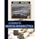
Ecografía Musculoesquelética
$455,000«Ecografía Musculoesquelética» aborda esta especialidad diagnóstica de forma total, para que sea de utilidad tanto a los que ya son especialistas en ecografía y quieren ampliar sus conocimientos y habilidades, como a los principiantes. Debido a que muchas de las dificultades del estudio de la ecografía musculoesquelética se deben a la incapacidad para evaluar las imágenes correctamente, hemos añadido en las ecografías de forma sistemática leyendas, referencias para sondas ecográficas, correlaciones uno a uno con fotografías clínicas, muestras anatómicas y quirúrgicas e imágenes obtenidas con otras técnicas. Además se han empleado muchos esquemas para enfatizar la descripción anatómica, los mecanismos y la biomecánica en el proceso de la enfermedad.
Stefano Bianchi, MD
Privat-docent, Université de Genève,
Consultant Radiologist,
Fondation et Clinique des Grangettes
Ginebra, SuizaCarlo Martinoli, MD
Associate Professor of Radiology,
Cattedra “R” di Radiologia –DICMI
Università di Genova
Genova, ItaliaEsta obra reúne en sus casi 1.000 páginas toda la información necesaria para la mejor identificación de las diferentes patologías musculoesqueléticas, su valoración clínica y su tratamiento. Los autores han diseñado unos originales y esquemáticos dibujos con los que se consigue dominar la anatomía ecográfica. A lo largo de todo el libro se correlacionan hallazgos ecográficos con TC y RM, mostrando, de cada técnica, tanto sus indicaciones como sus limitaciones.
Con los doctores Bianchi y Martinoli han colaborado diferentes especialistas, radiólogos, traumatólogos y reumatólogos, para obtener las más representativas imágenes y los análisis más certeros. El libro incluye muchas técnicas pioneras o mejoradas por los autores. El «Bianchi – Martinoli» es la obra más completa de su especialidad, establece un estándar de alta calidad en el área de la ecografía musculoesquelética y será considerado un clásico durante muchos años.
-
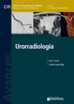
Avances en Diagnóstico por Imágenes: Urorradiología – Varela
$260,000Este nuevo volumen de la serie Avances en Diagnóstico por Imágenes sobre uroimágenes responde a los notables progresos realizados en las últimas décadas en relación con el conocimiento de la patología, la terapéutica y las técnicas de imagen, que se han convertido en una herramienta básica y fundamental en la toma de decisiones en la práctica urológica.
La posibilidad de evaluar con detalle la anatomía y patología urológica por imágenes, en particular con las nuevas técnicas de reconstrucción volumétrica, ha mejorado considerablemente la planificación y, por ende, la seguridad de la cirugía renal, urinaria y prostática. En el vol. Nº 8 de la serie, Retroperitoneo y páncreas, se abordaron ya los órganos retroperitoneales, y este nuevo libro complementa y enfatiza los avances urorradiológicos no solo en ese sector anatómico, sino también en el resto del sistema genitourinario. Se describe, en particular, la utilidad de las imágenes modernas en el manejo de la hematuria y la litiasis, así como en las patologías de la vía urinaria baja y órganos genitales. También se comentan detalles sobre terapéuticas mínimamente invasivas guiadas por imágenes, incluidos tratamientos recientemente introducidos en Latinoamérica como la crioablación de tumores y la embolización en hiperplasia benigna prostática. El libro se destaca por su diseño, el balance adecuado, la minuciosidad de los capítulos escritos didácticamente y una iconografía de alta
calidad, acorde con el nivel y seriedad habitual de los libros de la serie.$306,000 -
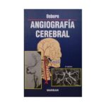
Angiografía Cerebral
Solicitar precioAl igual que sucede con todos mis libros, Angiografía Cerebral está concebido como un libro de enseñanza. He intentado unir, consolidar, simplificar e ilustrar la patología, epidemiología y, por supuesto, los hallazgos clínicos relativos a cada una de las principales enfermedades (y también de las menos importantes) que afectan a los vasos craneocerebrales. Para un repaso más fácil, los puntos clave se han resumido y destacado mediante cuadros que se incluyen en cada capítulo.
-
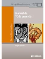
Manual de TC de urgencia + Libro electrónico – Ahualli
$236,000Manual de TC de urgencia es especialmente útil para el médico de guardia y el emergentólogo, ya que contiene conceptos de tomografía computarizada (generación de la imagen, tipos de tomógrafos, léxico adecuado, etc.), que permiten un entendimiento básico de esta importante herramienta diagnóstica. La mayor parte de la obra se destina a repasar aspectos generales y tomográficos de las principales patologías de presentación aguda (apendicitis aguda, peridiverticulitis aguda, litiasis ureteral, tromboembolismo pulmonar, disección aórtica, traumatismos, etc.), así como sus complicaciones más frecuentes (absceso periapendicular o peridiverticular, íleo biliar, necrosis pancreática, etc.). También se abordan procesos de evolución crónica, que en ocasiones pueden manifestar sintomatología aguda (tumores cerebrales, sinusitis e hipertrofia amigdalina, enfermedad de Crohn, etc.), así como ciertas entidades patológicas de urgencia, que si bien se manifiestan con poca frecuencia se caracterizan por presentar hallazgos tomográficos específicos (neumonía lipoidea exógena, bezoares del tracto gastrointestinal, etc.).
¿Cuándo hacer una TC, cómo y qué información ofrece? ¿Qué indican los hallazgos? Conciso y ampliamente ilustrado, Manual de TC de urgencias es la nueva obra de referencia para los radiólogos y emergentólogos.
$278,000 -
Manual de ecografía pediátrica – Gentile
$187,000Los estudios de imágenes en niños deben valorarse considerando las actuales normas de reducción de las dosis en radiaciones ionizantes. La ecografía se ha convertido en el método inicial de exploración por excelencia debido a que no emite radiaciones y a que numerosas enfermedades en los niños tienen demostración ecográfica.
En Manual de ecografía pediátrica, el Dr. Gentile transmite con precisión lo que él considera central para realizar un buen diagnóstico: Qué es lo que debe buscar el pediatra y qué le debe demostrar el ecografista. Para ello, reúne más de 1300 signos ecográficos en los que se describen, sintetizan y puntualizan los hallazgos de más de 350 patologías pediátricas, que se ilustran en 480 imágenes. El Anexo de medidas referenciales es un valioso aporte en el que se incluyen listados de diagnósticos diferenciales probables en relación al tamaño de los órganos. El manual resulta así un esfuerzo de recopilación y síntesis realizado por el autor a lo largo de su práctica profesional y docente, único en la bibliografía existente en idioma español.
Manual de ecografía pediátrica es una obra esencial para la formación de pediatras clínicos y especialistas, médicos generalistas, ecografistas y radiólogos, interesados en ecografía pediátrica.El Dr Fernando Gentile es Director de la carrera de posgrado de Diagnóstico por Imágenes pediátrico de la Universidad de Buenos Aires.
$220,000


Be the first to review “Pediatric Sonography”
You must be logged in to post a review.