-
Manual Practico de Medidas en Radiología Pediátrica – Montoya
$28,000En la práctica diaria de nuestra profesión y especialmente en el ejercicio de la Radiología Pediátrica, el radiólogo se ve en la necesidad de realizar medidas, muchas de ellas difíciles de encontrar y memorizar, poco claras y sometidas a una gran variabilidad dada por la edad y el sexo. Esta necesidad nos llevó a través de los años a recolectar un gran número de medidas que nos facilitarán y ayudarán en el diagnóstico.
Por medio de sencillos y prácticos diagramas de fácil consulta, el libro resume y explica de una forma didáctica, todas aquellas medidas necesarias en el ejercicio de la Radiología Pediátrica.
-
Radiology Review Manual
$865,000For more than 25 years, Dr. Dähnert’s Radiology Review Manual has earned its reputation as the green «bible” for board exam preparation, in teaching situations, and in the daily practice of radiology. A logical organization, extensive lists of image findings and differential diagnoses, an accessible outline format, and a thorough index have made this reference the #1 choice for success on the written boards. The Eighth Edition has been completely updated to provide head-to-heel coverage of the information needed for today’s general radiology practice.
NEW to this edition:
- A section on General Radiology has been created that covers techniques in nuclear medicine as well as contrast media, statistics, sedation, analgesia, and local anesthesia.
- Mnemonics have been added to help make need-to-know facts and trivia easier to find, review, and remember.
Your book purchase includes a complimentary download of the enhanced eBook for iOS, Android, PC & Mac. Take advantage of these practical features that will improve your eBook experience:
- The ability to download the eBook on multiple devices at one time — providing a seamless reading experience online or offline
- Powerful search tools and smart navigation cross-links that allow you to search within this book, or across your entire library of VitalSource eBooks
- Multiple viewing options that enable you to scale images and text to any size without losing page clarity as well as responsive design
- The ability to highlight text and add notes with one click
-
Manual de Radiología Mamaria
$222,000La radiología mamaria se ha convertido en la herramienta más eficaz para la detección precoz del cáncer de mama y la consecuente aplicación de la terapia más precisa y eficaz en el tratamiento de esta enfermedad. Sin embargo, los constantes avances en diagnóstico y terapia suponen un verdadero reto en la puesta al día de los diversos profesionales implicados. Esta obra es una guía multidisciplinar que pretende orientar al profesional ante diversas situaciones relacionadas con la patología mamaria.
Este manual está dividido en cinco secciones que abarcan las cuestiones principales acerca de la radiología de la mama:
- Sección I: profundiza en la visión multidisciplinar de la radiología mamaria, fundamental para poder entender y coordinar las actuaciones de profesionales de otros departamentos y servicios.
- Sección II: aborda las situaciones especiales que se encuentra el radiólogo, como el embarazo, lactancia, infancia, y adolescencia, en función de las características de los pacientes, así como de algunas estructuras anatómicas (como por ejemplo la axila) que merecen un enfoque diferenciado.
- Sección III: presenta los distintos protocolos de actuación de cara a la toma de decisiones, repasando las situaciones más cotidianas, como el manejo de la lesión palpable y no palpable, qué hacer ante una secreción mamaria, etc.
- Sección IV: muestra los manuales operativos de los procedimientos más habituales en las unidades radiológicas de mama, de manera detallada y diferenciada, según los niveles y tipos de responsabilidad de cada persona que en ellos intervienen.
- Sección V: revisa los criterios e indicadores de calidad de los procedimientos diagnósticos e intervencionistas, tanto desde el punto de vista técnico, como de cualificación profesional y de resultados.
$261,000 -
Avances en Diagnóstico por Imágenes: Cabeza y cuello
$245,000Los avances en el conocimiento de la patología de la cabeza y el cuello, región que integra la Neurorradiología junto con las patologías del sistema nervioso central y columna vertebral, nos motivaron a editar este nuevo volumen.
Con el fin de mantener la unidad editorial y la visión de conjunto, ofrecimos el cargo de Editor Invitado al Dr. Enrique Palacios Mayorga, médico neurorradiólogo de la Universidad de Tulane, a quien agradecemos haber aceptado hacerse cargo de esta tarea.
El contenido de Cabeza y Cuello se ha conformado con interesantes tópicos a cargo de autores cuya opinión es ampliamente reconocida por haberse destacado en su actividad profesional y académica en el campo de las neuroimágenes. Provenientes de diversos países (Canadá, Colombia, Estados Unidos, España, Panamá y México), estos profesionales actualizan el conocimiento y los avances sobre la anatomía de la cabeza y el cuello, la patología del plexo braquial, los senos paranasales y la órbita, y las neoplasias de cabeza y cuello, incluyendo laringe, adenopatías y base del cráneo. Asimismo, se incluyen conceptos diagnósticos sobre implantes dentales, sordera y alteraciones congénitas.
La importancia y actualidad de estos temas nos hacen confiar en que este noveno volumen, tal como los volúmenes precedentes de la Serie Avances en Diagnóstico por Imágenes, también será de utilidad en el desempeño profesional de los colegas y especialistas en formación. Por esta razón, agradecemos también al Dr. Palacios y a todos sus colaboradores por participar en este programa educativo que patrocina el Colegio Interamericano de Radiología.
-
Ecografía abdominal, aprendizaje paso a paso
$300,000Siguiendo el método del Dr. Berthold Block, usted aprenderá a realizar e interpretar la ecografía abdominal (vasos, hígado, hilio hepático, vesícula, páncreas, estómago, duodeno, diafragma, bazo, riñones, glándulas suprarrenales, vejiga, próstata y útero). El libro contiene:
Unidades didácticas claras.
Planos de abordaje sencillos y con orientación práctica para cada órgano.
Indicaciones sistemáticas para cada paso.
Más de 750 imágenes ecográficas y más de 250 esquemas explicativos.
Detallada descripción de todas las etapas del movimiento del transductor y de la producción de la imagen.
Esquemas en 3D para situaciones espaciales complejas.
Imágenes seriadas que facilitan la comprensión espacial.
Guías breves para el examen ecográfico de cada órgano: objetivos del estudio, cortes estándar, evaluación, trampas, etc.
Capítulo especial dedicado a documentación que incluye numerosas imágenes de referencia para hallazgos normales.Conviértase en un experto en las siguientes acciones:
Reconocer e interpretar todos los fenómenos ecográficos importantes.
Localizar y delimitar los órganos del abdomen.
Evaluar estructuras complejas.
Reconocer y diferenciar los hallazgos normales y anormales con seguridad.
Examinar de manera sistemática y completa los hallazgos casuales durante la consulta ecográfica.
Documentar los estudios de forma clara y completa.$353,000 -
Imagenología Mamaria, Guia práctica para un mejor diagnóstico
$570,000Incluye la nueva clasificación BI-RADS®
Un abordaje práctico basado en casos que le enseña a seleccionar y utilizar de manera óptima las opciones del diagnóstico por imágenes actual, para detectar e identificar en forma correcta las lesiones mamarias en el estadio más temprano posible.
» Aprenda a formular las preguntas correctas cuando observa una mamografía, una RM o una ecografía.
» Concéntrese en la información clave que debe conocer. Los capítulos concisos y organizados, seguidos de estudios de casos, reforzarán y ampliarán sus conocimientos.
» Distinga entre las variedades normales y las lesiones similares al cáncer gracias a la guía experta y clara en todos los métodos de diagnóstico por imágenes relevantes.
» Interprete los hallazgos más probables de ver en la práctica con la ayuda de imágenes de alta calidad, reforzadas con flechas y señalizaciones, que le ayudarán a reconocer e identificar lesiones sospechosas. -
Breast Imaging Companion
$360,000A pragmatic, common sense approach to the detection, evaluation and management of breast diseases and related imaging findings!
The fourth edition of this best selling “how-to” book includes major revisions, including the expansion of the screening mammography and breast MRI chapters, as well as the addition of digital breast tomosynthesis studies. Rather than having selected cropped images, the print and online versions of this book provide the reader with thousands of high quality images and complete imaging evaluations, from the screening images to the diagnostic mammogram, and—when appropriate—images from ultrasound, MRI, imaging guided biopsy, and preoperative wire localizations. Bulleted “key-facts” describe clinical, imaging and histological findings for a spectrum of breast diseases. With this book, breast-imaging radiologists are strongly encouraged to provide clinical, imaging and pathology concordance for optimal patient care, as well as direct and clinically relevant communication with providers and patients.
Key Features:
- Picture yourself in front of a screening mammogram or breast MRI… what now? How do you know if the study is interpretable? What are you looking for? Where do you look? If you detect something, what is the next appropriate step and how do you describe the finding?
- You will have access to hundreds of complete patient evaluations with thousands of images that include screening and diagnostic mammography, digital breast tomosynthesis, ultrasound, magnetic resonance studies and imaging guided breast related procedures with relevant pathology results and, when appropriate, the pathological stage.
- Develop appropriate differential considerations for the spectrum of breast imaging findings and appropriate management strategies.
- Review the indications for imaging guided procedures with step-by-step descriptions for each procedure illustrated with diagrams and images.
- Establish an optimal QA/QC program for your mammography practice, based on the concepts published by the ACR, regarding testing across digital platforms in the online version of the book.
- Test your knowledge and skills with a self-assessment chapter online.
Your book purchase includes a complimentary download of the enhanced eBook for iOS, Android, PC & Mac. Take advantage of these practical features that will improve your eBook experience:
- The ability to download the eBook on multiple devices at one time — providing a seamless reading experience online or offline
- Powerful search tools and smart navigation cross-links that allow you to search within this book, or across your entire library of VitalSource eBooks
- Multiple viewing options that enable you to scale images and text to any size without losing page clarity as well as responsive design
- The ability to highlight text and add notes with one click
-
Huesos y Articulaciones en imágenes radiológicas
Solicitar precioLas primeras incursiones de Resnick en práctica de la radiología fueron la entrega de informes de su padre (también radiólogo especializado en la imagen musculoesquelética) a los médicos que habían ordenado estudios de rayos X, por lo general para confirmar fracturas sospechosas. Sus mas de cuarenta años de experiencia en el mundo de la radiología avalan este clásico que continúa siendo el libro de consulta mas completo del mercado.
En la actualidad los métodos diagnósticos aplicados de forma rutinaria en el análisis de los trastornos musculoesqueléticos, incluyen no solo las radiografías convencionales, sino TC, RM, ecografías, gammagrafías y artografías. Estas técnicas son prácticamente las que aportan los nuevos contenidos y actualizan la tercera edición de este Best Seller» que contiene 4.000 imágenes.
-
Diagnóstico por imagenes del tórax pediátrico – Moenne
$241,000Publicada con gran éxito en 2005, la primera edición de este libro se convirtió en una de las obras de referencia en español para interpretar los estudios de imágenes del tórax pediátrico.
Esta segunda edición fue revisada en su totalidad por las autoras, quienes agregaron nuevas imágenes de gran interés para ilustrar las patologías. El libro cuenta con capítulos sobre anatomía del sistema respiratorio pediátrico, técnicas de imágenes y protección radiológica, tórax normal, infecciones respiratorias bajas, alteraciones de la vía aérea y malformaciones congénitas, entre otros. Además de la actualización de los temas, se abordan también las nuevas modalidades diagnósticas.$283,000 -
Manual de TC de urgencia + Libro electrónico – Ahualli
$236,000Manual de TC de urgencia es especialmente útil para el médico de guardia y el emergentólogo, ya que contiene conceptos de tomografía computarizada (generación de la imagen, tipos de tomógrafos, léxico adecuado, etc.), que permiten un entendimiento básico de esta importante herramienta diagnóstica. La mayor parte de la obra se destina a repasar aspectos generales y tomográficos de las principales patologías de presentación aguda (apendicitis aguda, peridiverticulitis aguda, litiasis ureteral, tromboembolismo pulmonar, disección aórtica, traumatismos, etc.), así como sus complicaciones más frecuentes (absceso periapendicular o peridiverticular, íleo biliar, necrosis pancreática, etc.). También se abordan procesos de evolución crónica, que en ocasiones pueden manifestar sintomatología aguda (tumores cerebrales, sinusitis e hipertrofia amigdalina, enfermedad de Crohn, etc.), así como ciertas entidades patológicas de urgencia, que si bien se manifiestan con poca frecuencia se caracterizan por presentar hallazgos tomográficos específicos (neumonía lipoidea exógena, bezoares del tracto gastrointestinal, etc.).
¿Cuándo hacer una TC, cómo y qué información ofrece? ¿Qué indican los hallazgos? Conciso y ampliamente ilustrado, Manual de TC de urgencias es la nueva obra de referencia para los radiólogos y emergentólogos.
$278,000 -
EL Sistema Bethesda para Informar la Citología Cervical – Nayar
$241,000La tercera edición ampliada y revisada de El Sistema Bethesda para informar la citología cervical ofrece información clara y actualizada sobre el modo de informar los hallazgos citológicos de las muestras cervicales, vaginales y anales conforme a la actualización del Sistema Bethesda 2014. En esta edición se incorporó un nuevo capítulo, se actualizaron la terminología y los contenidos, y se esclarecieron diferentes cuestiones terminológicas y morfológicas. Además, se incluyeron nuevas imágenes que reflejan la experiencia adquirida con la citología en medio líquido desde la publicación de la última edición en inglés (2004). De las más de 300 imágenes, algunas representan ejemplos clásicos de una entidad, mientras que otras ilustran dilemas de interpretación, características citomorfológicas limítrofes (borderline) o hallazgos que simulan anomalías epiteliales.
El Sistema Bethesda para informar la citología cervical es un libro de fácil consulta, indispensable para patólogos, citopatólogos, residentes de Patología, citotécnicos y ginecólogos.
$283,000 -
Stoller’s Orthopaedics and Sports Medicine: The Knee
$1,050,000View the knee like never before with this outstanding multimedia learning tool from a world-renowned expert! Stoller’s Orthopaedics and Sports Medicine: The Knee is now available and includes Stoller lecture videos, “Stoller Notes,” plus much more.
More than 25 years of trailblazing knee research and clinical experience are combined into one comprehensive, must-have edition. Now you can access this essential knowledge, along with the most extensive cadaver MR correlation available, to hone your skills in knee imaging. Concise, bulleted text accompanied by hundreds of clear line drawings, full-color illustrations, and high-quality images allow for rapid understanding and easy access to information.
Stoller’s Orthopaedics and Sports Medicine: The Knee includes:
- Print book
- 3.5 Hours of never-before-released videos of Dr. Stoller’s lecture slides with accompanying narration
- The full text in ebook format with enhanced navigation
- ‘Stoller Notes’ that provide deeper insight into the material
- A powerful search tool that pulls results from the book, reader’s notes, and the web
- Cross-linked PubMed pages, references, and more
- Note taking, highlighting, sharing and tabbing tools for easy reference to key content
This one-of-a-kind edition also offers:- One of the most comprehensive collections of color illustrations of orthopaedic pathoanatomy available
- The proven Stoller Checklist approach to MR joint interpretation for accurate and reproducible image interpretation
- Key concepts that emphasize and reinforce critical information
- Detailed and comprehensive figure legends that not only describe an image, but provide essential information about the disorder
- 3T and high resolution MR images
- Arthroscopy and surgical correlation
- New coverage of meniscal repair; isometry and graft position, plus double bundle; posterolateral corner; combined injury pattern, ACL plus PCL; patellar stabilization; and more
-
Imaging of the Athlete, An Issue of Radiologic Clinics of North America
$297,000This issue of Radiologic Clinics of North America focuses on Imaging of the Athlete, and is edited by Drs. Adam Zoga and Johannes Roedl. Articles will include: The Thrower’s Shoulder; Multimodality Imaging and Imaging Guided Therapy for the Painful Elbow; The Skeletally Immature and Newly Mature Throwing Athlete; Imaging Throwing Injuries Beyond the Shoulder and Elbow; Imaging Adductor Injury and «The Inguinal Disruption»; Image Guided Core Intervention and Postop Imaging; Core Injuries Remote from the Pubic Symphysis; MRI and MR Arthrography of the Hip; Knee Meniscus Biomechanics and Microinstability; Imaging Turf Toe and Traumatic Forefoot Injury; Imaging the Postoperative Knee; The Hindfoot Arch: What Role does the Imager Play?; Using Imaging to Determine Return to Play; and more!
Author Information
By Adam C. Zoga, MD, Assistant Professor of Radiology, Director of Musculoskeletal MRI and Ambulatory Imaging Services, Thomas Jefferson University Hospital, Philadelphia, PA and Johannes B. Roedl, MD, Thomas Jefferson University -
MRI of the Prostate
$390,000Although prostate cancer is the second leading cause of cancer death in men in the USA, it can be treated successfully if detected early. Disease management has gradually changed to a paradigm that relies on close monitoring through active surveillance in select patients, as well as ongoing refinements in treatment interventions, including minimally invasive procedures. This has resulted in a critical need for a more exacting methodology for performing targeted biopsies, assessing risk levels, and devising treatment strategies. Prostate MRI has emerged as the most precise, state-of-the-art imaging modality for prostate cancer diagnosis and management, thereby creating an immediate demand for radiologists to become proficient in its use.
Conceived and edited by a leading authority, with contributions from renowned experts in the field, MRI of the Prostate: A Practical Approach is the first book to tackle this important topic. It provides an overview of the fundamentals of prostate MRI acquisition, interpretation, and reporting. Readers will benefit from a wide range of insightful perspectives gleaned from years of hands-on experience.
Key Highlights
- Prostate Imaging Reporting and Data System (PI-RADS) for prostate MRI interpretation and cancer risk scoring
- Clinical pearls on the optimization and application of prostate MRI for risk assessment, disease staging, MRI-targeted biopsy, recurrent disease, and active surveillance
- The emerging utilization of PET and PET/MRI for primary prostate cancer evaluation
- More than 700 illustrations with one entirely image-based chapter featuring educational case studies
Radiologists will learn how to optimally perform and interpret prostate MRI, and referring physicians will learn to integrate it into day-to-day practice. This book is an essential resource for radiologists and radiology residents, as well as urologists, oncologists, MRI technicians, and other medical practitioners who treat patients with genitourinary disorders.
-
Diagnóstico en Patología: Tumores en Tejidos Blancos
$265,000El diagnóstico de los tumores de los tejidos blandos, suele plantear dificultades a los patólogos. Estos tumores no sólo son raros, sino que también adoptan una amplia gama de aspectos y, con frecuencia, las diferencias entre las entidades son sutiles.
Esta obra trata de aportar una información concisa y práctica sobre cada uno de los tumores, de ilustrar la gama de aspectos que se pueden encontrar al microscopio y de facilitar el diagnóstico diferencial, al permitir su comparación con otras lesiones parecidas.
En todos los capítulos el texto tiene un formato uniforme, en el que se incluyen rasgos clínicos, anatomopatológicos, inmunohistoquímicos, citogenéticos, de genética molecular en los casos en los que esta tiene importancia, y también una serie de diagnósticos diferenciales. Todos los diagnósticos se ilustran con imágenes clínicas, radiológicas y microscópicas que permiten valorar la amplia gama de aspectos y variaciones que se pueden encontrar. En las leyendas detalladas que acompañan a cada una de las imágenes, se incluyen otros puntos y claves para el diagnóstico. Los datos clave sobre cada entidad, se resumen en un recuadro distinto para poder consultarlo con rapidez, y en muchos capítulos la información sobre la inmunohistoquímica se presenta en forma de tabla para mayor comodidad.
El contenido se organiza en función de las líneas de diferenciación, siguiendo la clasificación de consenso de la OMS, aunque añadiendo algunos tumores no incluidos en la misma, como las neoplasias neurales y cutáneas, y una serie de entidades de más reciente caracterización. Para tratar de dar respuesta a la creciente demanda por parte de los patólogos y de otros médicos de capturar los datos en una forma sinóptica o estructurada, hemos añadido unos protocolos convencionales para el estudio e informe de los sarcomas de tejidos blandos.
Basta con revisar la bibliografía, para apreciar que nuestros conocimientos acerca de la anatomía patológica de los tumores de tejidos blandos, están aumentando a una sorprendente velocidad. Se describen entidades nuevas, algunas de las conocidas se están revaluando en estudios clinicopatológicos y están surgiendo nuevas técnicas de investigación y opciones terapéuticas dependientes de la anatomía patológica.
Esperamos que Diagnóstico en Patología – Tumores en tejidos blandos» resulte útil en la práctica diaria, para facilitar el diagnóstico, el informe y, por supuesto, el tratamiento.
-
Diagnóstico en Patología: Hepatobiliar y Pancreático
$236,000A menudo, la patología hepática clínica supone un reto para los patólogos , porque puede existir un notable solapamiento entre las entidades de cualquier diagnóstico diferencial concreto. Las lesiones neoplásicas del hígado, el páncreas y las vías biliares también pueden ser difíciles de diagnosticar, especialmente si no son hallazgos frecuentes. Diagnóstico en Patología: hepatobiliar y pancreática está diseñado para ayudar tanto a los patólogos en prácticas como en ejercicio, a enfrentarse a estos retos.
Si una imagen vale más que mil palabras, este trabajo contiene al menos 1.295.000 palabras en forma de imágenes. Estas incluyen fotomicrografías, fotografías macroscópicas y preciosas ilustraciones médicas, muchas de las cuales han sido creadas especialmente para este libro.
Como en los restantes libros de la colección «Diagnóstico en Patología», las datos clave relativos a cada diagnóstico aparecen resaltados en un cuadro para facilitar su uso. En la sección «Diagnóstico diferencial», se presenta una lista de entidades importantes para cada diagnóstico. Además, se incluyen protocolos de estadificación estructurados para todas las neoplasias malignas, así como tablas con las inmunotinciones pertinentes y capítulos sobre la disección macroscópica de las piezas quirúrgicas.
En nombre de los autores de este libro, esperamos que Diagnóstico en Patología: hepatobiliar y pancreática» se convierta en el libro de referencia favorito, al cual recurran una y otra vez los patólogos en su práctica diaria.
-
Diagnóstico en Patología: Genitourinario
$364,000Los casos de patología urológica, constituyen una proporción significativa de los que podemos ver en cualquier departamento de Anatomía Patológica. Si hablamos de los nuevos casos que se presentan cada año, el carcinoma de próstata es el proceso maligno no cutáneo más frecuente, y los carcinomas vesicales y renales, o de la pelvis renal, ocupan el cuarto y séptimo lugar.
En los últimos años, se ha trabajado mucho para aclarar los aspectos clinicopatológicos de estos y otros cánceres urológicos, y es esencial actualizar nuestros conocimientos para mantener el papel central que ocupa la patología en el diagnóstico y el tratamiento de dichos pacientes.
«Diagnóstico en Patología – Genitourinario» pretende ser un compendio para el diagnóstico «en un solo paso», de fácil manejo para el patólogo ocupado, para los residentes y para los especialistas en formación.
La combinación de un texto conciso, con los datos en forma de viñetas y las diversas ilustraciones, ayudan al patólogo a llegar al diagnóstico correcto, a considerar las principales posibilidades de diagnóstico diferencial, utilizar cuando proceda los estudios complementarios y seguir las normas pertinentes para elaborar informes de anatomía patológica completos y precisos. Las numerosas ilustraciones representan no solo las presentaciones morfológicas clásicas, sino también las menos frecuentes, y todas ellas se acompañan de leyendas con aspectos formativos que realzan el texto. No solo existe una amplia cobertura de las entidades más frecuentes, sino también de aquellas que no lo son tanto, o que incluso son extraordinariamente raras como los tumores renales pediátricos, los carcinomas asociados a translocación de la familia MiTF/TFE, el carcinoma basaloide y adenoide quístico de la próstata, los tumores del estroma de los cordones testiculares, el adenocarcinoma de la red testicular y el carcinoma epidermoide seudohiperplásico o el carcinoma cuniculatum del pene.
El libro se ha organizado en 5 secciones: Tumores y afecciones de tipo tumoral del riñón, Vejiga urinaria, Próstata y vesículas seminales, Testículos y estructuras paratesticulares y Pene y Escroto. Al final de cada sección se incluye el protocolo para la elaboración de informes de muestras resecadas, así como tablas y paneles inmunohistoquímicos exhaustivos para el diagnóstico diferencial.
-
Simulacros de Examen 5: Nervioso II
$30,000«Simulacros de examen» es una colección de cuadernos para el entrenamiento y la autoevaluación en anatomía humana.
Cada cuaderno consta de dos partes, la primera es una serie de láminas de autoevaluación compatibles con cualquier atlas anatómico y la segunda es una batería de preguntas tipo test con respuesta.
Volumen 5 : Nervioso II
-
Simulacros de Examen 4: Nervioso I
$20,000«Simulacros de examen» es una colección de cuadernos para el entrenamiento y la autoevaluación en anatomía humana.
Cada cuaderno consta de dos partes, la primera es una serie de láminas de autoevaluación compatibles con cualquier atlas anatómico y la segunda es una batería de preguntas tipo test con respuesta.
Volumen 4 : Nervioso I
$25,000 -
Simulacros de Examen 3: Articulaciones
$30,000Simulacros de examen» es una colección de cuadernos para el entrenamiento y la autoevaluación en anatomía humana.
Cada cuaderno consta de dos partes, la primera es una serie de láminas de autoevaluación compatibles con cualquier atlas anatómico y la segunda es una batería de preguntas tipo test con respuesta.
Volumen 3 : Articulaciones
-

Simulacros de Examen 2: Huesos
$30,000«Simulacros de examen\» es una colección de cuadernos para el entrenamiento y la autoevaluación en anatomía humana. Cada cuaderno consta de dos partes, la primera es una serie de láminas de autoevaluación compatibles con cualquier atlas anatómico y la segunda es una batería de preguntas tipo test con respuesta.
Volumen 2 : Huesos
-
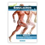
Simulacros de examen 1: Músculos
$30,000«Simulacros de examen» es una colección de cuadernos para el entrenamiento y la autoevaluación en anatomía humana. Cada cuaderno consta de dos partes, la primera es una serie de láminas de autoevaluación compatibles con cualquier atlas anatómico y la segunda es una batería de preguntas tipo test con respuesta.
Volumen 1 : Músculos
-
-
Evidencias en Cardiología VIII (2Vol)
$248,000Cuando hace algunos años decidimos confluir en lo que luego se consolidó como GEDIC -Grupo de Estudio, Docencia, e investigación Clínica- nos animaba la idea de generar un ámbito de intercambio creativo en el análisis y desarrollo de la investigación clínica, como así también en la docencia de su metodología. La organización, durante dos años consecutivos, del curso de Cardiología Basada en la Evidencia, nos permitió convocar a un grupo de cardiólogos jóvenes de excelente formación y con vocación para revisar sistemáticamente la información disponible y cuestionar (a veces, hasta obsesivamente) las bases que fundamentan nuestras conductas cotidianas en el paciente concreto. De una maduración de los materiales utilizados para el curso surgió como un desarrollo inmanente la concreción de la primera edición de este libro, que ha sumado luego la entusiasta coloboración de nuevos coautores. El libro se ha expandido y multiplicado de un pequeño manual a una obra voluminosa, y alcanza hoy su octava edición.
Este libro no hubiera sido posible sin la colaboración incondicional de todos los coautores, que se han mantenido y acrecentado a los largo de los años, y la incansable tarea de los coordinadores, comenzando por Raúl Schwartzman, y luego Daniel Ferrante, Juan Gagliardi, Javier Mariani, Maximiliano de Abreu, Ignacio de Urquiza y en esta edición Natalia Vensentini.
Mantenemos como modalidad nueva de lectura la actualización de la app Evi-Cardio (r) para smartphones y tablets, que incluye más de mil ensayos clínicos resumidos con una modalidad que permite su análisis individual, efectuar metaanálisis sencillos, y consultar los materiales fundamentales del libro. La actualización de estos materiales a través de la vía electrónica nos permitirá mantener con los lectores un contacto más activo en los próximos años.
Hemos contado con la erudita colaboración del Dr. José Tessler, codirector de GEDIC, que ha hecho mucho para que nuestros conceptos estadísticos maduraran y se ampliarana. Su pérdida irreparable nos compromete aún más en difundir su clara visión científica y ética.
El Dr. Hernán Doval quiere agradecer especialmente al Dr. Raúl Oliveri y al Hospital Italiano por la oportunidad de aprender y practicar la cardiología en un ambiente de camaradería, libre discusión científica y responsabilidad en las decisiones médicas. Y a los pacientes, que con sus interesantes y diversas historias de vida, nos hacen médicos eficientes.
El Dr. Carlos Tajer quiere a su vez agradecer a sus grandes maestros en la Cardiología, los Dres. Raúl Oliveri y Carlos Bertolasi, que le han brindado su conocimiento, apoyo y marcado un rumbo ético en la profesión y a su esposa e hijos por su afecto y colaboración en los largos fines de semana, noches y madrugadas invertidos en esta obra.
Ambos agradecen a los lectores que han impulsado esta obra y la han transformado en un material de consulta y debate vivo en la práctica de la cardiología.
$331,000 -
Valvulopatías
$370,000Escrito por destacados cardiólogos y cirujanos cardiovasculares, el libro describe las valvulopatías más frecuentes e importantes en todos sus aspectos clásicos: etiopatogenia, fisiopatología, anatomía patológica, semiología, evaluación clínica e historia natural, así como los tratamientos médicos, endovasculares y quirúrgicos. Con una visión actual e integradora, aporta valiosa información sobre la toma de decisiones, controversias y consensos que permite profundizar el entendimiento de las alternativas que ofrece actualmente la cardiología para la elección de prótesis cardíacas, el tratamiento de pacientes con estenosis aórtica por reemplazo valvular aórtico, la aplicación de técnicas de resincronización, el uso de la técnica de Ross, entre otros.
Los puntos sobresalientes y los conceptos insoslayables se enumeran al comienzo de cada capítulo a modo de una breve guía para abordar cada tema. Ampliamente ilustrado con imágenes a color, Valvulopatías incluye un acceso web a videos con imágenes diagnósticas de la más alta calidad, que permitirán al lector familiarizarse con los hallazgos propios de cada patología.
Incluye acceso web a videos y material suplementario.$435,000