
-
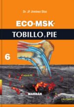
Eco MSK 6 Tobillo y Pie – Jiménez
$316,000En este nuevo libro ECO•MSK, Tobillo y Pie se hace un completo recorrido de la anatomía ecográfica, la patología y las técnicas intervencionistas de esta región. Por una parte, se realiza una revisión ampliada de la sonoanatomía de la pierna y del tobillo, estudiando con mucho detalle algunas estructuras como los ligamentos de la articulación. Tal es el caso de los fascículos de la cara posterior del tobillo o los ligamentos cortos que unen los huesos del pie, que se explican con gran precisión.
Pero lo más novedoso es la somera descripción que se realiza de la anatomía ecográfica de las estructuras de la planta del pie, incluyendo los músculos intrínsecos de la región plantar, los tendones de los músculos extrínsecos como el tendón del peroneo largo, el nudo de Henry y el tendón tibial posterior. Además, se evalúan las articulaciones metatarsofalángicas y los haces neurovasculares.
En cuanto al examen ultrasonográfico de la patología, se revisan las lesiones musculares de la pierna y las lesiones articulares, tendinosas y ligamentosas del tobillo. También es novedoso el amplio repaso que se realiza de las lesiones que afectan a los huesos, articulaciones y tendones del pie. Pero lo que mas destaca es el estudio en profundidad de aquellas afecciones específicas del pie, como las lesiones de la fascia, los neuromas, las lesiones quísticas o la metatarsalgia, además de las lesiones que afectan a la piel y al tejido celular subcutáneo. Finalmente se describen las técnicas mas frecuentes de intervencionismo ecoguiado aplicadas a estas lesiones de pie y tobillo.
$372,000 -
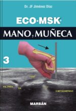
Eco Msk 3 Mano Y Muñeca – Jiménez
$271,000La muñeca es otro ejemplo de la utilidad de la herramienta ecográfica que se ha incorporado a la práctica diaria de numerosas especialidades médicas para el diagnóstico y tratamiento ecodirigido de las lesiones que en la muñeca y mano se producen.
En ECO•MSK MANO y MUÑECA se hace un completo recorrido por la anatomía ecográfica de esta región, describiendo las pequeñas articulaciones, así como aquellas zonas no descritas en anteriores textos como la división del nervio cubital dentro del canal de Guyon, la descripción de los músculos de la región volar de la mano o la organización de los tendones flexores en su recorrido a través de los dedos.
Además, se revisan con detalle las lesiones de esta región anatómica, estudiando los diferentes procedimientos ecoguiados que se aplican para la resolución de estas patologías. Se incluye por tanto el examen de las lesiones tendinosas centradas en los procesos tenosinoviales y roturas de los tendones que cruzan la muñeca y la mano. También se describe el examen ecográfico de los síndromes de compresión neurológica, las lesiones óseas, articulares y ligamentosas, los quistes y tumores y, finalmente, las lesiones que afectan específicamente a los dedos de la mano.
Un ejemplo de la utilidad de esta técnica en la evaluación del síndrome del túnel carpiano es su capacidad para detectar las alteraciones morfológicas del nervio, secundarias a los procesos de compresión en el túnel y correlacionarlas con la gravedad del daño nervioso en todos los casos, incluso en los idiopáticos. Además, permite por una parte identificar la mayoría de las formas asociadas que provocan este síndrome, y por otra resaltar algunas variantes o condiciones anatómicas predisponentes que pueden representar contraindicaciones para tratamientos mínimamente invasivos, además de evaluar a aquellos pacientes con un resultado desfavorable después de un tratamiento quirúrgico.
Por todo ello, en este volumen, hemos conseguido un triple objetivo. Reunir toda la información necesaria para alcanzar un amplio conocimiento de la sonoanatomía de la mano y de la muñeca, así como de los signos ecográficos de las lesiones que allí se producen y, finalmente, de los procedimientos ecodirigidos para el tratamiento de la patología que con más frecuencia en esta región anatómica se origina.
$319,000 -
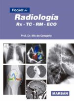
Pocket de Radiología RX – TC – RM – ECO – De Gregorio
$164,000¡Pocket de Radiología es el libro que contempla todas las técnicas (RM, TC, RX, Eco) en formato de bolsillo!
Moderno, práctico y didáctico, distinguiéndose por «no sólo ser» para radiólogos. Es el compañero ideal para una guardia para cualquier médico general o residente que, de una forma rápida, podrá aclarar cualquier duda.
Desde la desaparición, hace ya muchos años, del «Compendio de Pedrosa», no había habido ningún libro que cubriera esa demanda y Pocket de Radiología ofrece la posibilidad de utilizar su contenido como libro de consulta en un formato Pocket, tremendamente cómodo de manejar y con una excelente calidad de imagen, con fotografías radiológicas mejoradas digitalmente e impresas
$192,000 -
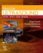
Vascular Ultrasound How, Why and When, 4th Edition – Thrush
$333,000Now in its fourth edition, Vascular Ultrasound offers a compact yet comprehensive practical guide for anyone working in the field of vascular sonography.
The book is written by expert practitioners as an easily accessible reference, providing key information suited to sonographers in their day-to-day practice. It covers essential vascular investigations undertaken by ultrasound departments and vascular laboratories in more detail than general ultrasound textbooks, but without overwhelming sonographers with highly complex information that may not be relevant to them.
Here you will find essential information including the principle of ultrasound physics to enable accurate assessment of the peripheral circulation and blood flow, the use of the main scanner functions and controls, the main disorders of the arterial and venous circulation system with appropriate treatment and management, and techniques for the diagnosis and grading of disease.
-
Neuroimaging Anatomy, Part 2: Head, Neck, and Spine – Massoud
$480,000In this issue of Neuroimaging Clinics, guest editor Dr. Tarik F. Massoud brings his considerable expertise to the topic of Neuroimaging Anatomy, Part 2: Head, Neck, and Spine. Anatomical knowledge is critical to reducing both overdiagnosis and misdiagnosis in neuroimaging. This issue is part two of a two-part series on neuroimaging anatomy that focuses on the head, neck, and spine. Each article addresses a specific area such as the orbits, sinonasal cavity, temporal bone, pharynx, larynx, and spinal cord.Key Features- Contains 14 relevant, practice-oriented topics including anatomy of the orbits; maxillofacial skeleton and facial anatomy; temporal bone anatomy; craniocervical junction and cervical spine anatomy; anatomy of the spinal cord, coverings, and nerves; and more.
- Provides in-depth clinical reviews on neuroimaging anatomy of the head, neck, and spine, offering actionable insights for clinical practice.
- Presents the latest information on this timely, focused topic under the leadership of experienced editors in the field. Authors synthesize and distill the latest research and practice guidelines to create clinically significant, topic-based reviews.
-
Neuroimaging Anatomy, Part 1: Brain and Skull, An Issue of Neuroimaging Clinics – Massoud
$480,000In this issue of Neuroimaging Clinics, guest editor Dr. Tarik F. Massoud brings his considerable expertise to the topic of Neuroimaging Anatomy, Part 1: Brain and Skull. Anatomical knowledge is critical to reducing both overdiagnosis and misdiagnosis in neuroimaging. This issue is part one of a two-part series on neuroimaging anatomy that focuses on the brain, with each article addressing a specific area. The issue also includes an article on Brain Connectomics: the study of the brain’s structural and functional connections between cells.Key Features- Contains 13 relevant, practice-oriented topics including anatomy of cerebral cortex, lobes, and the cerebellum; brainstem anatomy; cranial nerves anatomy; brain functional imaging anatomy; imaging of normal brain aging; and more.
- Provides in-depth clinical reviews on neuroimaging anatomy of the brain and skull, offering actionable insights for clinical practice.
- Presents the latest information on this timely, focused topic under the leadership of experienced editors in the field. Authors synthesize and distill the latest research and practice guidelines to create clinically significant, topic-based reviews.
-
Joe / Breast Imaging, 4th Ed. The Core Requisites
$520,000Focusing on high-yield information, Breast Imaging: The Core Requisites, 4th Edition emphasizes the basics to help you establish a foundational understanding of breast imaging during rotations, refresh your knowledge of key concepts, and learn strategies to provide «value-added» reports to referring clinicians. This completely rewritten and reorganized edition emphasizes the essential knowledge you need in an easy-to-read format, with thorough updates that cover new imaging modalities, the latest guidelines, and integration of physics information throughout.
Key Features-
- Emphasizes the essentials in a templated, quick-reference format that includes numerous outlines, tables, pearls, boxed material, and bulleted content for easy reading, reference, and recall.
-
- Helps you build and solidify core knowledge to prepare you for clinical practice with critical, up-to-date information on mammography, breast ultrasound, digital breast tomosynthesis, and breast MRIs, as well as special chapters on lymph node evaluation in breast imaging, augmented and reconstructed breast, and special populations in breast imaging.
-
- Features hundreds of high-quality images, including correlations of ultrasound, mammography, digital breast tomosynthesis and MRI.
-
- Published as part of the newly reimagined Core Requisites series, an update to the popular Requisites series aimed at radiology trainees and today’s busy clinicians.
-
- An eBook version is included with purchase. The eBook allows you to access all of the text, figures and references, with the ability to search, customize your content, make notes and highlights, and have content read aloud.
-
-
The Physics of Clinical MR Taught Through Images 5th – Runge
$498,000The objective of this 5th edition of the book, as with the prior editions, is to teach through images a practical approach to magnetic resonance (MR) physics and image quality. Unlike other texts covering this topic, the focus is on clinical images rather than equations. A practical approach to MR physics is developed through images, emphasizing knowledge of fundamentals important to achieve high image quality. Pulse diagrams are also included, which many at first find difficult to understand. Readers are encouraged to glance at these as they go through the text. With time and repetition, as a reader progresses through the book, the value of these and the knowledge thus available will become evident (and the diagrams themselves easier to understand).
$540,000 -
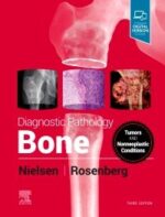
Diagnostic Pathology: Bone, 3rd Edition – Nielsen
$1,580,000This expert volume in the Diagnostic Pathology series is an excellent point-of-care resource for practitioners at all levels of experience and training. Covering all areas of bone pathology, it incorporates the most recent clinical, pathological, and molecular knowledge in the field to provide a comprehensive overview of all key issues relevant to today’s practice. Richly illustrated and easy to use, Diagnostic Pathology: Bone is a one-stop reference for accurate, complete surgical pathology reports, ideal as a day-to-day reference or as a reliable training resource.
-
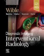
Diagnostic Imaging: Interventional Radiology, 3rd Edition – Wible
$1,606,000Key Features• Provides a comprehensive, expert reference for review and preparation of common and infrequently performed procedures, with detailed «step-by-step» instructions for conducting image-guided interventions in various clinical scenarios
• Covers vascular venous, arterial, and lymphatic procedures, with specific attention to thromboembolic, posttransplant, and oncologic therapies
• Addresses emerging nonvascular image-guided treatments in pain management, neurologic and musculoskeletal procedures, and others
• Contains new procedures chapters on endovascular treatments for pulmonary embolisms and deep vein thrombosis, prostate artery embolization, pelvic venous disorders, and percutaneous/endovascular arteriovenous fistula (AVF) creation
• Features sweeping updates throughout, including updated guidelines and recommendations from the Society of Interventional Radiology
• Offers more than 3,200 images (in print and online), including radiologic
images, full-color medical illustrations, instructional photo essays, and clinical
and histologic photographs
• Clearly demonstrates procedural steps, complications, treatment alternatives, variant anatomy, and more?all fully annotated to highlight the most important diagnostic information
• Organized by procedure type, allowing for quick comparison of different procedural techniques that may have complementary or alternative roles in managing specific disease states
• Builds on the award-winning second edition, which won first prize in the British Medical Association’s Medical Book Awards, Radiology category
• Includes the enhanced eBook version, which allows you to search all text, figures, and references on a variety of devices
$1,785,000 -
Chest Radiology: A Resident’s Manual – Kirchner
$391,000Chest Radiology: A Resident’s Manual is a comprehensive introduction to reading and analyzing radiologic cardiopulmonary images. Readers are guided through systemic image analysis and can further enhance their learning experience with training cases found at the end of each chapter. Cases describe and discuss frequently asked questions regarding heart failure, bronchitis, pneumonia, bronchial carcinoma, fibrosis, pleural disorders, and more. This user-friendly manual will allow the reader to confidently answer the most important and commonly encountered questions related to plain chest radiographs in daily clinical practice. The easy-to-read layout pairs explanatory text on the left page with related drawings and images on the right, allowing readers to navigate their way through each section with ease. Features More than 600 high-resolution images and illustrations demonstrate a wealth of pathology Concise descriptions explain how to examine conventional x-ray and CT images Numerous callout boxes in each chapter highlight key takeaway points A scratch-off code provides access to a searchable online database of 250 must-know thoracic imaging cases This practice-oriented manual is an invaluable resource and reference guide for residents and radiologists-in-training.
$460,000 -
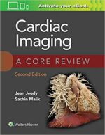
Cardiac Imaging: A Core Review – Jeudy
$350,000Prepare for success on the cardiac imaging component of the radiology Core Exam! Cardiac Imaging: A Core Review, 2nd Edition, by Drs. Jean Jeudy and Sachin Malik, is an up-to-date, practical review tool written specifically for the Core Exam. This helpful resource contains 300 image-rich, multiple-choice questions with detailed explanations of right and wrong answers, fully revised content, high-yield tables for easy review, and additional eBook questions to ensure you’re ready for the Core Exam or recertification exam.
- Features questions in all exam areas, including basic imaging, normal anatomy, all diseases relative to cardiac imaging, all modalities, and postoperative appearances of devices.
- Features over 200 high-resolution images.
- Provides concise answers with explanations of each choice followed by relevant, up-to-date references.
- Follows the structure and content of what you’ll encounter on the test, conveniently organized by topic.
Enrich Your eBook Reading Experience
Read directly on your preferred device(s), such as computer, tablet, or smartphone.
Easily convert to audiobook, powering your content with natural language text-to-speech.
-
Diagnostic Imaging: Head and Neck, 4th Edition – Hamilton
$1,529,000Covering the entire spectrum of this fast-changing field, Diagnostic Imaging: Head and Neck, fourth edition, is an invaluable resource for neuroradiologists, general radiologists, and trainees—anyone who requires an easily accessible, highly visual reference on today’s head and neck imaging. Dr. Philip R. Chapman and his team of highly regarded experts provide up-to-date information on recent advances in disease identification, imaging techniques, and tumor staging to help you make informed decisions at the point of care. The text is lavishly illustrated, delineated, and referenced, making it a useful learning tool as well as a handy reference for daily practice.$1,699,000 -
Diagnostic Imaging: Genitourinary – Fananapazir
$1,611,000Covering the entire spectrum of this fast-changing field, Diagnostic Imaging: Genitourinary, fourth edition, is an invaluable resource for general radiologists and trainees—anyone who requires an easily accessible, highly visual reference on today’s genitourinary (GU) imaging. Drs. Ghaneh Fananapazir, Bryan R. Foster, and their team of highly regarded experts provide up-to-date information on recent advances in technology and the understanding of GU diseases and disorders to help you make informed decisions at the point of care. The text is lavishly illustrated, delineated, and referenced, making it a useful learning tool as well as a handy reference for daily practice.Key Features- Serves as a one-stop resource for key concepts and information on GU imaging, including a wealth of new material and content updates throughout
- Features more than 2,500 images (state-of-the-art cross-sectional imaging, full-color medical illustrations, radiologic images, clinical photographs, H&E stains, and gross pathology photographs), plus 500 additional images and video clips online
- Features updates from cover to cover including updated Bosniak 2019 criteria, PI-RADS v2.1 terminology, updated cancer staging chapters, new interventional techniques, new contrast agent guidelines, and new chapters on transgender imaging
- Contains a new 10-chapter section on kidney transplant, including post-transplant procedures
- Covers key procedures such as renal biopsy; percutaneous genitourinary interventions; kidney ablation/embolization; and venous sampling and venography
- Covers all aspects of GU imaging, including typical and variant findings; GU anatomy, physiology, imaging protocols, and work-ups; and new developments in diagnostic criteria and terminology
- Uses bulleted, succinct text and highly templated chapters for quick comprehension of essential information at the point of care
- Enhanced eBook version included with purchase. Your enhanced eBook allows you to access all of the text, figures, and references from the book on a variety of devices
$1,790,000 -
Problem Solving in Pediatric Imaging – Sarvis
$840,000Optimize diagnostic accuracy with Problem Solving in Pediatric Imaging, a new volume in the Problem Solving in Radiology series. This concise title offers quick, authoritative guidance from experienced radiologists who focus on the problematic conditions you’re likely to see—and how to reach an accurate diagnosis in an efficient manner.Key Features- Addresses the practical aspects of pediatric imaging—perfect for practitioners, fellows, and senior level residents who may or may not specialize in pediatric radiology, but need to use and understand it.
- Integrates problem-solving techniques throughout, addressing questions such as, «If I see this, what do I need to consider? What are my next steps?»
- Presents content in a highly useful, real-world manner, with sections on conventional radiography in the ED, NICU, PICU, and CICU; fluoroscopy; body imaging; and neuroradiology.
- Imaging findings are merged with clinical, anatomic, developmental, and molecular information to extract key diagnostic and therapeutic information.
- Contains a section on special topics with chapters on radiation safety and quality assurance.
- Features hundreds of high-quality color images and anatomic drawings that provide a clear picture of what to look for when interpreting studies. Illustrations conveying normal anatomy help you gain an in-depth perspective of each pathology.
- Enhanced eBook version included with purchase. Your enhanced eBook allows you to access all of the text, figures, and references from the book on a variety of devices.
-
Manual de Ecografía Musculoesquelética – Ventura
$98,000Este manual tiene como objetivo de servir como guía para los Reumatólogos que se entrenan en ultrasonido musculoesquelético, cubre aspectos fundamentales como los principios físicos de la ecografía, el conocimiento de los elementos de un ecógrafo para obtener imágenes óptimas, la revisión de las estructuras articulares y periarticulares, la revisión sistemática de las regiones anatómicas, la utilidad del Doppler de poder, los hallazgos sonográficos en las diferentes patologías en las que el ultrasonido ha demostrado su utilidad tanto para el diagnóstico como para seguimiento, además del papel del ultrasonido en patologías menos frecuentes como vasculitis, afección de glándulas salivales, lesiones traumáticas y, por último pero no menos importante, el uso del ultrasonido como guía para punciones-infiltraciones. Cuenta con la participación de renombrados Reumatólogos con gran experiencia en la técnica, que apoyan a ECOMER (Escuela de Ecografía del Colegio Mexicano de Reumatología).
-
Diagnóstico por imágenes del sistema musculoesquelético – Kimura
$201,000La serie de libros del Dr. Kenji Kimura ofrece una selección de los casos clínicos radiológicos recopilados durante sus más de 40 años de práctica profesional. Los casos se encuentran distribuidos al azar —como ocurre en la práctica diaria— y presentan diferentes grados de dificultad: desde los más sencillos hasta los que requieren de experiencia y sagacidad para descubrir signos y hallazgos sutiles a la hora de arribar a un diagnóstico. En cada volumen se presentan inicialmente las imágenes y los datos clínicos claves de cada caso, lo que permite al lector ensayar un diagnóstico presuntivo. Posteriormente, se ofrece el diagnóstico final acompañado de una discusión minuciosa, documentada con bibliografía actualizada. Se trata de una serie de lectura ágil y amena, y con un enorme valor didáctico. Diagnóstico por imágenes del sistema musculoesquelético es el tercer volumen de la serie. Aborda la patología ósea, articular, muscular y de los tejidos blandos, que incluyen ligamentos y tendones, con el uso coordinado de una variedad de técnicas de imagen, que han evolucionado por el desarrollo de nuevas tecnologías. Está dirigido a estudiantes de medicina, residentes de radiología, traumatólogos, ortopedistas, internistas, radiólogos y especialistas en resonancia magnética y ultrasonido del sistema musculoesquelético.
$237,000 -
Tc de Cuerpo Serie Secretos – Strang
$149,000Ediciones Journal publica en espanol esta obra de la afamada coleccion Secretos de la editorial Elsevier, que ofrece pautas para realizar e interpretar correctamente la tomografia computarizada de cuerpo. Estructurada en base a 1469 preguntas y respuestas, junto a 696 imagenes de TC, el lector encontrara las caracteristicas distintivas de los titulos anteriores de la serie: listas, reglas nemotecnicas y consejos de los autores. Este nuevo volumen contiene otras mejoras que le resultaran de gran utilidad:* Un nuevo diseno en dos colores, recuadros con Conceptos fundamentales y listas de sitios de Internet para consultar: Un tamano mas pequeno y comodo, Un capitulo inicial que resume los 100 secretos principales de la TC de cuerpo. Cualquiera sea la pregunta que surja acerca de la TC de cuerpo, esta obra tiene todas las respuestas.
$184,000 -
Radiología Básica «Método programado para el aprendizaje» FORA
$279,000La Radiología ha pasado de ser una disciplina auxiliar en la Medicina en el siglo XX a ser la base de la mayor parte de las decisiones clínicas en el siglo XXI. Prueba de ello es que, el peso de la especialidad ha ido incrementando en las últimas convocatorias del examen MIR. A pesar de ello, la mayoría de los planes de estudios de las universidades siguen manteniendo a la Radiología en un discreto segundo plano.
Al igual hace unos años, la publicación de Radiología Esencial permitió proporcionar un texto de referencia a la formación de los residentes de Radiodiagnóstico, la sección de Formación en radiología de la SERAM presenta este texto, adaptado al estudiante de Medicina, con el fin de llenar un hueco no resuelto en la formación del médico general.
Esta obra aborda todas las habilidades y conocimientos que un alumno debe adquirir durante su formación, incluidas las relacionadas con aspectos técnicos, de radioprotección y de las distintas técnicas de imagen radiológica, así como de medicina nuclear. Para ello, los capítulos están acompañados de recursos didácticos como textos destacados, casos clínicos, preguntas de autoevaluación y multitud de figuras necesarias para la comprensión de la materia.
$328,000 -
Diagnostic Imaging: Pediatrics, 4th Edition
$1,699,000Covering the entire spectrum of this fast-changing field, Diagnostic Imaging: Pediatrics, fourth edition, is an invaluable resource for pediatric radiologists, general radiologists, and trainees—anyone who requires an easily accessible, highly visual reference on today’s pediatric imaging. Dr. A. Carlson Merrow, Jr., and his team of highly regarded experts provide up-to-date information on recent advances in technology and safety in the imaging of children to help you make informed decisions at the point of care. The text is lavishly illustrated, delineated, and referenced, making it a useful learning tool as well as a handy reference for daily practice.Key Features- Serves as a one-stop resource for key concepts and information on pediatric imaging, including a wealth of new material and content updates on more than 400 diagnoses
- Features more than 2,500 illustrations including radiologic images, full-color illustrations, endoscopic and bronchoscopic photographs, clinical photos, and gross pathology images
- Features updates from cover to cover including specifics from revised disease classifications and new terminology in best practices recommendations for radiologic reporting
- Reflects evolving imaging technology in conjunction with increased awareness of radiation, contrast, and anesthesia safety in children, and how these advances continue to alter pediatric imaging approaches
- Uses bulleted, succinct text and highly templated chapters for quick comprehension of essential information at the point of care
- Enhanced eBook version included with purchase. Your enhanced eBook allows you to access all of the text, figures, and references from the book on a variety of devices
-
Diagnostic Imaging Musculoskeletal Non-Traumatic Disease, 3rd Edition – Davis
$1,790,000Covering the entire spectrum of this fast-changing field, Diagnostic Imaging: Musculoskeletal Non-Traumatic Disease, third edition, is an invaluable resource for musculoskeletal radiologists, general radiologists, and trainees—anyone who requires an easily accessible, highly visual reference in this complex area of imaging. Drs. Kirkland W. Davis, Donna G. Blankenbaker, Stephanie A. Bernard, and their team of highly regarded experts provide up-to-date information on recent advances in technology and the understanding of musculoskeletal diseases and disorders to help you make informed decisions at the point of care. The text is lavishly illustrated, delineated, and referenced, making it a useful learning tool as well as a handy reference for daily practice.Key Features-
- Guides readers through the complexities of the full range of non-traumatic musculoskeletal disorders, including arthritis, collagen vascular diseases, bone tumors, soft tissue tumors, infections, systemic diseases, developmental and congenital abnormalities, and metabolic diseases
-
- Contains multiple new chapters on topics such as musculoskeletal genetics, neurinomas, and rapidly progressive osteoarthritis, among others, as well as updates throughout on reclassified lesions, tumors, and neoplasms; musculoskeletal infection details, including image-guided aspirations and biopsies for infections; and evolving medical and surgical treatments for many musculoskeletal conditions
-
- Reflects recent changes in the World Health Organization’s classification of tumors and tumor-like conditions regarding terminology and diagnostic criteria
-
- Covers evolving imaging techniques such as ultrasound in non-traumatic disease imaging, contrast-enhanced ultrasound use in tumor biopsies, enhanced MR of musculoskeletal tumors, and diffusion-weighted MR, and PET/CT and PET/MR use for rapidly progressive osteoarthritis
-
- Provides up-to-date discussions of enhancements in bone and soft tissue tumor pathology and imaging of orthopedic implants and related hardware
-
- Features more than 3,750 annotated images (with an additional 2,100+ digital-only examples), including radiologic images, full-color medical illustrations, clinical and histologic photographs, and gross pathology images
- Uses bulleted, succinct text and highly templated chapters for quick comprehension of essential information at the point of care
-
-
Bontrager Manual de posiciones y técnicas radiológicas – Lampignano
$150,000Mediante listas de instrucciones y fotografías de pacientes en las posiciones correctas, esta guía de bolsillo le ayudará a colocar a los pacientes con seguridad, rapidez y confianza durante los estudios radiográficos más frecuentes. Este manual de posiciones y anatomía radiológicas presenta también técnicas y criterios de valoración que le permitirán evaluar rápida y fácilmente las radiografías que realice en su práctica clínica. Como novedades presenta fotografías actualizadas que muestran la tecnología digital más reciente en radiología, con nuevas imágenes radiográficas, de posiciones y de equipos; incorpora las proyecciones de Bernageau y Zanca, indicadas en la patología y los traumatismos de hombro y 217 proyecciones resumen la información esencial. Asimismo, incluye contenido actualizado que recoge las últimas competencias y directrices establecidas por el ARRT Asimismo, las presentaciones sobre las posiciones contienen instrucciones sobre la posición, el tamaño del campo de colimación, la colocación y el ángulo del RC, y los intervalos de kVp propuestos, y se deja espacio para anotar los factores de exposición.
$176,000 -
SEUS Ecografía Musculoesquelética Esencial
$207,000Los equipos actuales de ecografía de alta resolución permiten obtener imágenes de las partes blandas con una gran definición, lo que junto a su inocuidad, portabilidad y capacidad de examen dinámico y comparativo, convierten a este método en una herramienta fundamental para el estudio de la patología del aparato locomotor y para guiar la realización de diversos procedimientos terapéuticos. Sin embargo, su eficacia está condicionada a las características del equipo y a la capacitación del operador, por lo que resulta fundamental una sólida formación teórica y práctica para dar la respuesta adecuada a la creciente demanda y variedad de exploraciones.
La amplia experiencia asistencial y docente de todos los autores queda recogida con precisión, orden y amenidad en los temas tratados en este libro que, tras un primer capítulo de conceptos teóricos generales, está estructurado por áreas anatómicas con una metodología basada en la anatomía ecográfica, la sistemática de exploración y las patologías más frecuentes. El texto está ilustrado con numerosos gráficos e imágenes representativas.
Una excelente obra que nace con el espíritu de transformarse en un libro básico de cabecera. Su clara finalidad docente y su orientación práctica facilitarán el estudio de todos los profesionales de la salud interesados en la ecografía músculo-esquelética y sus contenidos serán de gran ayuda a la hora de resolver las dudas que a menudo se plantean durante la realización de las exploraciones ecográficas.
$243,000 -
Atlas de Bolsillo de Cortes Anatómicos Tomo 2. Tomografía computarizada y resonancia magnética: tórax, corazón, abdomen y pelvis – Moller
$169,000Este atlas de bolsillo, claro y fácil de usar, es reconocido por sus magníficas ilustraciones y la capacidad de representar la anatomía seccional en cada plano. Con sus dos tomos acompañantes, constituye una herramienta de navegación altamente especializada para todos los clínicos que necesitan dominar la anatomía radiológica e interpretar minuciosamente las imágenes por RM y TC.
Características especiales del Atlas de bolsillo de Cortes Anatómicos:
- Organización didáctica en unidades de dos páginas, con imágenes radiológicas de alta calidad en una cara y excelentes dibujos, a todo color, en la otra.
- Cientos de imágenes de RM y TC en alta resolución, muchas obtenidas mediante la última generación de escáneres (p. ej., RM 3T, TC de 64 cortes).
- Código de color unificado, que facilita la identificación de estructuras similares en los diferentes cortes.
- Leyendas concisas y fáciles de leer en todas las imágenes.
Actualizaciones de la 4ª edición del Tomo II:
- Imágenes por TC del tórax y el abdomen en los tres planos: axial, sagital y coronal.
- Nueva cubierta desplegable en la solapa de contraportada con los segmentos pulmonares y hepáticos y las estaciones ganglionares.
- Seguimiento de las clasificaciones internacionales estandarizadas de la American Heart Association para los vasos cardíacos y del AJCC/UICC para los ganglios linfáticos mediastínicos.
Compacto, fácil de usar, muy visual y diseñado para la búsqueda rápida, este libro es ideal para su uso tanto en el entorno académico como clínico.
$199,000 -
Atlas de Bolsillo de Cortes Anatómicos Tomo 1. Tomografía computarizada y resonancia magnética: cabeza y cuello – Moller
$169,000Este atlas de bolsillo, claro y fácil de usar, es reconocido por sus magníficas ilustraciones y la capacidad de representar la anatomía seccional en cada plano. Con sus dos tomos acompañantes, constituye una herramienta de navegación altamente especializada para todos los clínicos que necesitan dominar la anatomía radiológica e interpretar minuciosamente las imágenes por RM y TC.
Características especiales del Atlas de bolsillo de Cortes Anatómicos:
- Organización didáctica en unidades de dos páginas, con imágenes radiológicas de alta calidad en una cara y excelentes dibujos, a todo color, en la otra.
- Cientos de imágenes de RM y TC en alta resolución, muchas obtenidas mediante la última generación de escáneres (p. ej., RM 3T, TC de 64 cortes).
- Código de color unificado, que facilita la identificación de estructuras similares en los diferentes cortes.
- Leyendas concisas y fáciles de leer en todas las imágenes.
Actualizaciones de la 4ª edición del Tomo I:
- Nuevas secuencias de imágenes craneales axiales y coronales del hueso temporal mediante TC.
- Sección de RM aumentada con todas las imágenes 3T RM nuevas del lóbulo temporal e hipocampo, la arteria basilar, los nervios craneales y el seno cavernoso, entre otras estructuras.
- Nuevas secuencias de angiografía arterial por RM del cuello e imágenes adicionales de la laringe.
Compacto, fácil de usar, muy visual y diseñado para la búsqueda rápida, este libro es ideal para su uso tanto en el entorno académico como clínico.
$199,000


