
-
Wortsman / Dermatologic Ultrasound with Clinical and Histologic Correlations Hardcover
$998,000Significant technological advances have produced equipment that allows imaging of the skin with variable frequency ultrasound in previously unseen detail and provides a range of dynamic data that is currently unmatched by any other technology. Dermatologic Ultrasound with Clinical and Histologic Correlations is a comprehensive introduction to ultrasonography of the skin, nails, and scalp as it relates to the assessment and diagnosis of dermatologic diseases. It provides radiologists, sonographers, dermatologists, and physicians with interest in skin imaging with a concise understanding of the diagnosis of dermatologic conditions through extensive high-resolution gray scale and color Doppler ultrasound images and presents classical correlations of clinical dermatologic lesions with sonographic and histologic findings. Featuring more than 1700 images, this text-atlas provides an excellent starting point in learning about this topic.
-
Diagnóstico por imágenes del hombro E-Book
$108,000Libro Electrónico
En Diagnóstico por Imágenes del hombro, el Dr. Burgos Flor aborda de manera exhaustiva el examen del hombro normal y patológico, utilizando múltiples modalidades de imágenes que permiten el estudio específico de esta articulación. La obra desarrolla temas tales como las enfermedades articulares, traumatismos, tumores óseos y de los tejidos blandos, síndrome del manguito rotador y patología del bíceps braquial y bursal, inestabilidad del hombro, atrapamiento nervioso y articulación esternoclavicular.
Con más de 500 imágenes e ilustraciones de excelente calidad, Diagnóstico por Imágenes del hombro es un libro práctico y de lectura necesaria para radiólogos, ecografistas, técnicos radiólogos, traumatólogos y cirujanos de hombro. -
Manual de técnicas intervencionistas guiadas por imágenes E-Book
$248,000Auspiciado por la Sociedad Iberoamericana de Intervencionismo (SIDI), este libro en el que participan reconocidos
especialistas de la región, tiene por objeto poner a disposición de todos los profesionales de América Latina
un material útil, práctico y actualizado sobre las diferentes técnicas intervencionistas. Además de describir las
diferentes técnicas, sus indicaciones y resultados, se incluyen sugerencias, consejos y detalles que suelen encontrarse
en la práctica diaria. El Manual de técnicas intervencionistas guiadas por imágenes será el único en
su tipo en idioma español, y contribuirá a la difusión de las técnicas intervencionistas mínimamente invasivas,
tanto para médicos en formación como para los profesionales de la salud interesados en esta especialidad y sus
diferentes procedimientos, y a las aplicaciones de la técnica en un amplio espectro de especialidades. -
MR Imaging of the Prostate, An Issue of Radiologic Clinics of North America
Solicitar precioThis issue of Radiologic Clinics of North America focuses on MR Imaging of the Prostate, and is edited by Dr. Aytekin Oto. Articles will include: Prostate cancer diagnosis and management: A urologist’s perspective; MRI of prostate zonal anatomy; Technique of Multi-parametric MRI of the prostate; Multi-parametric MRI- interpretation including PIRADS v2; Prostate MRI for screening and active surveillance; Prostate MRI for staging; Prostate MRI for post-treatment evaluation and recurrence; Pitfalls in prostate MRI; MRI-targeted prostate biopsies; MRI-guided focal treatment for prostate cancer; Role of Prostate MRI in radiation oncology; Challenges and future directions of prostate MRI; and more!
-
Emergent Neuroimaging: A Patient Focused Approach: Neuroimaging Clinics
Solicitar precioThis issue of Neuroimaging Clinics of North America focuses on Emergent Neuroimaging: A Patient Focused Approach, and is edited by Dr. Diego B. Nunez. Articles will include: The adult patient with acute neurologic deficit: an update on imaging trends; The patient with thunderclap headache; Imaging considerations in the patient with intracerebral hemorrhage; Emergent neuroimaging in the oncologic and immunosuppressed patient; Emergent neuroimaging during pregnancy and puerperium; Imaging the unconscious “found down” patient in the ED; Neurological emergencies in the pediatric patient including accidental and non-accidental trauma; Head and neck injuries: special considerations in the elderly patient; Current challenges in the use of CT and MRI in suspected cervical spine trauma; Radiation considerations in emergent neuroimaging; Blunt cranio-cervical trauma: Does the patient have a cerebral vascular injury?; New and emerging patient-centered CT imaging and image-guided treatment paradigms for maxillofacial trauma; Acute neurological syndromes beyond stroke: The role of emergent MRI; and more!
-
Cardiovascular Magnetic Resonance
Solicitar precioWritten by an expert team of cardiologists, radiologists, and basic scientists, this third edition of Cardiovascular Magnetic Resonance continues to bridge the divide among specialty areas in with cohesive presentation of this complex and fast-changing field. Offering comprehensive coverage of CMR and the latest cardiology applications, this practical reference enhances the understanding of cardiac physiology and the interpretation and diagnosis of cardiovascular disease. This is an ideal resource for cardiologists, cardiovascular and general radiologists, and anyone who needs up-to-date information on CMR’s uses, benefits, and limitations in cardiovascular care.
Key Features
- Provides state-of-the-art coverage of CMR technologies and guidelines, including basic principles, imaging techniques, ischemic heart disease, right ventricular and congenital heart disease, vascular and pericardium conditions, and functional cardiovascular disease.
- Includes new chapters on non-cardiac pathology, pacemaker safety, economics of CMR, and guidelines as well as new coverage of myocarditis and its diagnosis and assessment of prognosis by cardiovascular magnetic resonance, and the use of PET/CMR imaging of the heart, especially in sarcoidosis.
- Features more than 1,100 high-quality images representing today’s CMR imaging.
- Covers T1, T2 and ECV mapping, as well as T2* imaging in iron overload, which has been shown to save lives in patients with thalassaemia major.
- Discusses the cost-effectiveness of CMR.
- Expert Consult™ eBook version included with purchase. This enhanced eBook experience allows you to search all of the text, figures, and references from the book on a variety of devices.
-
Colección CIR 17 E-Books + Libro Físico Tórax 2
$1,140,000Serie Avances en diagnóstico por imágenes
Los 16 volúmenes que conforman la colección CIR ahora en en formato epub 3.
Este formato permite una excelente visualización del contenido del libro en cualquier tamaño de pantalla.
Cada código permite el acceso y la descarga del libro en 2 dispositivos móviles y 2 de escritorio.
-
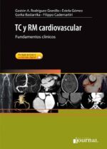
TC y RM Cardiovascular
$610,000Los avances tecnológicos en tomografía computarizada (TC) multicorte y resonancia magnética (RM) han permitido que, hoy en día, estas técnicas se hayan convertido en herramientas esenciales para el diagnóstico de las enfermedades cardiovasculares. Lejos de tratarse de técnicas de imagen que se utilizan cuando métodos más establecidos presentan limitaciones, en la actualidad se considera que el valor diagnóstico, pronóstico y de planificación terapéutica de estas modalidades excede su mero empleo como técnicas diagnósticas alternativas.
El objetivo de este libro es describir los fundamentos clínicos de la TC y la RM cardíaca y vascular, y contribuir a la necesaria unión que debe existir entre los distintos especialistas para lograr una visión más integral de este grupo de enfermedades. Incluye capítulos sobre aspectos fisiopatológicos de la enfermedad cardiovascular, dosis de radiación y tipos de material de contraste. Nutrido de un gran número de figuras en color, tablas y algoritmos diagnósticos, además de un amplio material complementario on line a modo de casos clínicos, permitirá aprender los secretos de la imagen cardiovascular no invasiva a cualquier profesional interesado en las particularidades de la imagen cardíaca y vascular, desde el cirujano cardíaco o vascular a los médicos clínicos especialistas, además del personal técnico y de enfermería. Con objeto de que sea un libro eminentemente práctico, su diseño facilita una lectura sencilla y comprensible que ayuda a asimilar los conocimientos con gran facilidad.
$718,000 -
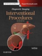
Diagnostic Imaging: Interventional Procedures
Solicitar precioMore than 100 interventional procedures, lavishly illustrated with 800+ outstanding medical images, highlight the second edition of this practical reference. Dr. Brandt C. Wible and his expert author team provide carefully updated information in a concise, bulleted format, keeping you current with recent advances in interventional radiology. Succinct text, outstanding illustrations, and up-to-date content make this title a must-have reference for trainees as well as seasoned interventionalists and vascular surgeons who need a single, go-to guide in this fast-changing area.
New to this Edition
-
- Meticulously updated throughout, with new information on interventional oncology, including radioembolizaiton, transarterial chemoembolization, and percutaneous ablation; IVC filter placement and removal; stroke intervention; and venous recanalization and thrombolysis
-
- Hundreds of high-quality case images and graphics (many new to this edition) clearly demonstrate procedural steps, complications, treatment alternatives, variant anatomy, and more-all fully annotated to highlight the most important diagnostic information
-
- New chapters including lumbar puncture and myelogram and celiac plexus block
-
- Newly streamlined discussions of procedural steps create a simpler, more focused text designed for quick reference
-
- Updated expected outcomes from recent prominent literature
- Expert Consult™ eBook version included with purchase. This enhanced eBook experience allows you to search all of the text, figures, Q&As, and references from the book on a variety of devices.
-
-
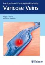
Varicose Veins
Solicitar precioThis is a compact, readable, highly usable, valuable guide to have on varicose veins and related conditions, including an overview of the human venous system, information pertaining to causative factors, diagnoses, symptoms, and treatment options… — BIZ INDIA
Chronic venous disease is a common condition, with a prevalence as high as 50% in industrialized countries worldwide. Of those, about 20-25% of women and 10-15% of men have visible varicose veins. Varicose vein treatment has become an increasingly multidisciplinary field, and one that has seen cutting-edge advances and significant growth.
Felipe B. Collares, Salomao Faintuch, and a team of venous disease experts have compiled, Varicose Veins, a concise book that covers the full range of interventional procedures for venous insufficiency. Following introductory chapters on anatomy and pathophysiology, the authors guide readers through the clinical exam, imaging, compression therapy, and various minimally invasive techniques. Highly practical and an affordable alternative to larger published tomes.
Key Highlights
- Step-by-step guide on core venous interventions – from compression therapy to sclerotherapy – ambulatory phlebectomy to endovascular ablation
- Illustrations delineate anatomy and specific treatment modalities
- Clinical pearls on patient safety and preventing complications
- Discussion of emerging endovascular approaches that do not require administration of tumescent anesthesia
This handy resource is a must-have for trainees and veteran physicians. The practical and focused layout provides a well-rounded reference for all specialists who perform varicose vein procedures – interventional radiologists, vascular surgeons, phlebologists, cardiologists, and dermatologists.
-
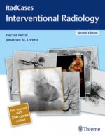
Radcases Interventional Radiology
$220,000Interventional Radiology Second Edition by Hector Ferral and Jonathan Lorenz details image-guided procedures and prepares you to diagnose the full range of arterial, venous, nonvascular, and oncological conditions. Included in this book are 100 cases covering congenital malformations to acquired pathologies, illustrated with clear, high-quality images and now with questions and answers for review. For maximum ease of self-assessment, each case begins with the clinical presentation on the right-hand page; study that and then turn the page for imaging findings, differential diagnoses with the definitive diagnosis, essential facts, pearls and pitfalls, and more.
Key Highlights
- 500 high-resolution images guide the reader through the cases
- The importance of the RV/LV ratio in CT evaluation of pulmonary embolism
- Cutting-edge advances in prostate embolization, thermal ablation applications, the use of IVC filters, and the management of vascular malformations and portal hypertension
- Arterial interventions with tips for aggressive management of below the knee arterial complications
- Evaluation of AAA stent grafts and common types of endoleaks, with tips on how to identify and describe them
-
Primer of Diagnostic Imaging
Solicitar precioWidely known as THE survival guide for radiology residents, fellows, and junior faculty, the “purple book” provides comprehensive, up-to-date coverage of diagnostic imaging in an easy-to-read, bulleted format. Focusing on the core information you need for learning and practice, this portable resource combines the full range of diagnostic imaging applications with the latest imaging modalities, making it the perfect clinical companion and review tool.
Key Features
- Features more than 1,200 detailed illustrations now in full color, plus images that clearly depict the latest applications of CT, MRI, PET/CT, and other diagnostic imaging modalities.
- Provides new coverage of non-interpretive skills such as quality and safe dosing.
- Balances new information and anatomic drawings with timeless, relevant material to fully prepare you for the boards and for daily practice.
- Explains the nuances of key diagnostic details for all body systems, including signs and symptoms, anatomic landmarks, and common radiologic-pathologic alterations, for the full range of radiologic modalities and specialties.
- Uses a bulleted format and provides mnemonics, descriptive terminology, and space for note taking that make it easy to learn and remember key facts, techniques, and images.
- Allows you to work through diagnoses with hundreds of differentials for board certification preparation.
- Clarifies the impact of the latest disease entities on the interpretation of radiologic findings.
- Expert Consult™ eBook version included with purchase. This enhanced eBook experience allows you to search all of the text, figures, and references from the book on a variety of devices.
-
Decision Making in Neurovascular Disease
Solicitar precioNeurovascular medicine has emerged as an established, semi-independent subspecialty of neurology and neurosurgery. Decision Making in Neurovascular Disease focuses on the challenging process of determining the best approach for managing patients with intracranial atherosclerosis, carotid artery disease, stroke, aneurysms, arteriovenous malformations, arteriovenous fistulae, cavernous malformations, and hypervascular tumors. Leonardo Rangel-Castilla, Robert Spetzler, esteemed coauthors, and an impressive cadre of experts discuss highly divergent modalities including medical management, open cerebrovascular, endovascular, radiosurgery, and combined/multimodality alternatives.
The book is organized into seven sections: Ischemic Stroke and Vascular Insufficiency, Aneurysms – Anterior Circulation, Aneurysms – Posterior Circulation, Aneurysms – Other, Arteriovenous Malformations and Fistula, Cavernous Malformations, and Hypervascular Tumors. Chapters include an introduction, decision-making algorithm, whether to treat, conservative management, anatomical considerations, clinical and imaging evaluation, differential diagnosis, treatment options, images, clinical and radiographic follow-up, and suggested reading.
Key highlights
- Simple algorithms accompanying 71 chapters supported by the latest, most updated information in the literature
- More than 300 radiologic images help elucidate disease-specific treatment decision making
- Step-by-step guidance, clinical pearls, surgical nuances, complication avoidance, and evidence-based outcomes provide in-depth understanding
- Point/counterpoint expert commentary on each case provides balanced insights on potential implications of specific treatments
This essential step-by-step book is a must-have for residents and fellows in neurosurgery, neurology, endovascular, interventional radiology, vascular neurology, and neurocritical care, as well as veteran clinicians in these specialties.
-
Ultrasound: A Core Review
$245,000Uniquely designed for the Core Exam, Ultrasound: A Core Review covers all key aspects of ultrasound, mimicking the image-rich, multiple-choice format of the actual test. Ideal for residents getting ready for the Core Examination, as well as practitioners taking recertification exams, this one-of-a-kind review follows the structure and content of what you’ll encounter on the test, effectively preparing you for Core Exam success!Key Features:
- Contains 300 image-rich ultrasound questions with answers, explanations, and references.
- Features high-quality black-and-white, color, and spectral Doppler images throughout.
- Covers organ-specific ultrasound as well as gynecologic, pregnancy, vascular, musculoskeletal, peritoneal space, retroperitoneum, abdominal wall, chest, and ultrasound-guided procedures.
- Includes questions on noninterpretive skills such as ultrasound physics and quality and safety.
- Provides answers and rationales as to why an answer is correct and other choices are incorrect.
Your book purchase includes a complimentary download of the enhanced eBook for iOS, Android, PC & Mac.
Take advantage of these practical features that will improve your eBook experience:
- The ability to download the eBook on multiple devices at one time — providing a seamless reading experience online or offline
- Powerful search tools and smart navigation cross-links that allow you to search within this book, or across your entire library of VitalSource eBooks
- Multiple viewing options that enable you to scale images and text to any size without losing page clarity as well as responsive design
- The ability to highlight text and add notes with one click
-
Abbreviated MRI of the Breast
$390,000Although mammography is the primary method used for breast cancer screening, screening mammography is limited especially in women with dense breasts, which includes nearly 50% of all women in the United States. Despite improvements such as digital mammography, computed aided detection, and digital breast tomosynthesis, breast cancer continues to be a leading cause of cancer-related death in women. The recent proliferation of screening breast ultrasound has led to increased health care costs and false positives, with only a slight improvement in breast cancer detection. It is time for a better test.
This is the first textbook dedicated to the subject of abbreviated breast MRI (AB-MR). The editors are principal investigators in the first multicenter trial evaluating AB-MR. Each chapter is authored by a leading expert in the field of breast MRI.
AB-MR only takes 10 minutes or less to perform, has a comparable cost to screening breast ultrasound, and detects twice as many cancers compared to combined screening with mammography and ultrasound. The improved performance of AB-MR is irrespective of breast density, family history, overall breast cancer risk, and cancer characteristics (e.g. type, staging, invasive or intraductal, primary or recurrent). As such, it will likely become a routine screening tool in women with dense breasts.
Key Features
- A background on breast MR imaging including a review of current research data
- Fundamental guidelines for implementing, performing, and interpreting AB-MR
- Technical approaches with proven efficacy, including biopsy methods
- Accurate interpretation presented in an easy-to-read flow chart format
- More than 250 high quality color illustrations
AB-MR has the potential to help radiologists overcome breast cancer screening limitations and change current standards of practice. This book provides radiologists with the necessary tools to quickly incorporate AB-MR into clinical practice, with an ultimate goal of improved breast cancer detection rates and patient outcomes.
-
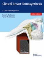
Clinical Breast Tomosynthesis – Salkowski
$555,000Digital breast tomosynthesis (DBT), popularly called “3D mammography” is a state-of-the-art breast imaging technology that provides the ability to view inside the breast, layer by layer. Advantages of DBT include elimination of superimposed tissue, improved detection of otherwise hidden lesions, and significantly higher cancer detection rates compared to conventional mammography.
Although it is increasingly being implemented in imaging centers, hands-on clinical learning tools have been lacking – until now. Written by renowned breast imaging specialists, this multimedia textbook is the most definitive DBT reference available. The first section is dedicated to DBT fundamentals, including physics, reconstruction methods, acquisition parameters, and patient dose and data size considerations. The next two sections are case-based clinical practice presentations, organized by DBT screening and diagnostic components.
Key Features
- The use of 3D breast imaging to detect and differentiate malignancies from normal and benign findings
- 100 case studies include pertinent clinical information, introductory images, and findings labeled on subsequent images with BI-RADS terminology
- Full field digital mammography and/or synthetic 2D mammogram images included in all case studies mirror what is typically encountered in DBT breast-imaging programs
- Clinical pearls on navigating the technology, interpreting images, making diagnoses, and decisions regarding follow-up studies
- Accompanying online videos further elucidate the still images
This remarkable, interactive reference is a must-have for trainee and practicing radiologists and will also benefit mammography technicians. The detailed case studies and videos demonstrate the full utility of DBT as a screening and diagnostic technology in context with mammography, ultrasound, and MRI of the breast.
$652,000 -

Caffey’s Pediatric Diagnostic Imaging, 2-Volume Set
Solicitar precioFor more than 70 years, Caffey’s Pediatric Diagnostic Imaging has been the comprehensive, go-to reference that radiologists have relied upon for dependable coverage of all aspects of pediatric imaging. In the 13th Edition, Dr. Brian Coley leads a team of experts to bring you up to date with today’s practice standards in radiation effects and safety, as well as in head and neck, neurologic, thoracic, cardiac, gastrointestinal, genitourinary, and musculoskeletal pediatric imaging. This two-volume bestselling reference is a must-have resource for pediatric radiologists, general radiologists, pediatric subspecialists, pediatricians, hospitals, and more – anywhere clinicians need to ensure safe, effective, and up-to-date imaging of children.
New to this Edition
-
- Takes an updated, contemporary approach with more focused and consistently formatted content throughout. Clinical content includes Overview; Etiologies, Pathophysiology, and Clinical Presentation; Imaging, including pros and cons, costs, evidence-based data, findings, and differential diagnostic considerations; and Treatment, including follow-up.
- Features 8,500 high-quality images – 1,000 new or updated.
-
- Provides expanded coverage of advanced imaging and diagnostics, including genetics and fetal imaging, MRI and advanced MR techniques, low-dose CT, ultrasound, nuclear medicine, and molecular imaging, as well as the latest quality standards, evidence-based data, and practice guidelines.
- Features new Key Points boxes and more tables and flowcharts that make reference faster and easier.
-
- Focuses on safety, particularly in radiation dosing, as part of the Image Gently® campaign to improve pediatric imaging while limiting radiation exposure and unneeded studies.
- Expert Consult™ eBook version included with purchase. This enhanced eBook experience allows you to search all of the text, figures, and references from the book on a variety of devices.
Key Features
- Provides access to 50 online videos, including hypertrophic pyloric stenosis, disorders of swallowing, fetal swallowing, fetal bowel obstruction, upper GI and ultrasound evaluation of malrotation and volvulus, congenital heart disease MRI evaluation, and many more.
- Includes separate chapters on radiation effects and safety, pre-natal imaging, neoplasms, trauma, techniques, embryology, genetic anomalies, and common acquired conditions.
-
-
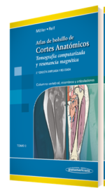
Atlas de Bolsillo de Cortes Anatómicos Tomo 3. Tomografía computarizada y resonancia magnética: Columna vertebral, Miembros y Articulaciones – Moller
$169,000Este atlas de bolsillo, claro y fácil de usar, es reconocido por sus magníficas ilustraciones y la capacidad de representar la anatomía seccional en cada plano. Con sus dos tomos acompañantes, constituye una herramienta de navegación altamente especializada para todos los clínicos que necesitan dominar la anatomía radiológica e interpretar minuciosamente las imágenes por RM y TC.
Actualizaciones de la 2ª edición del Tomo III:
- Agregado de 137 páginas y 234 nuevas ilustraciones y un total de 725 imágenes de gran calidad.
- Adición, a las imágenes centradas en las regiones cercanas a las articulaciones, de nuevas ilustraciones en dos planos de todo el brazo, el antebrazo, el muslo y la pierna.
- Descripción de las lesiones óseas o de los tejidos blandos de la diáfisis, como las causadas por la inflamación o por tumores.
$199,000 -
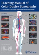
Teaching Manual of Color Duplex Sonography
Solicitar precioThe Third Edition of this up-to-date and user friendly workbook helps medical students, sonographers, residents, and radiologists gain a fundamental grasp of the application of color duplex ultrasound by reviewing normal findings, important pathologic conditions, scanning techniques, and the role and importance of color duplex ultrasound in detecting and assessing various disease states.
Topics include:
- Basic physical and technical principles
- Innovative techniques and ultrasound contrast agents (e.g., power Doppler, SieScape imaging, clarify vascular enhancement, tissue Doppler, precision upsampling, arterial stiffness, eTracking)
- Vascular surgery: peripheral arterial occlusive disease, venous insufficiency and thrombosis, AV fistulae, and aneurysms
- Endocrinology: thyroid gland
- Internal medicine: abdominal organs, lymph nodes, TIPSS
- Nephrology: kidneys and renal allografts
- Neurology: intra- and extracranial cerebral arteries
- Cardiology: B- and M-mode imaging, cardiac anomalies, wall motion analysis
- Urology: testicular torsion, tumors, erectile dysfunction
- Obstetrics and gynecology: tumors, anomalies, fetal perfusion defects
-
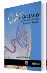
El Encéfalo Diagnóstico por imagen, patología y anatomía
$635,000La obra de la Dra. Anne G. Osborn, ‘Osborn, el encéfalo. Diagnóstico por imagen, patología y anatomía’ es completa, atractiva visualmente y fácil de entender. Proporciona un sólido y amplio marco para comprender una materia compleja y árida como es la imagen cerebral.
Combina los aspectos anatómicos esenciales con la patología macroscópica y las técnicas de imagen, mostrando de forma clara por qué y cómo aparecen las enfermedades de la forma en la que lo hacen.
Todo el texto ha sido redactado por la Dra. Osborn, aportándole su toque genuino y especial para convertir temas complejos en fácilmente comprensibles y dándole un enfoque uniforme a todos los capítulos que integran la obra.
Cubre todos los aspectos de obligado conocimiento en lo relativo a la imagen cerebral, junto con espectaculares ejemplos patológicos, la anatomía más relevante y técnicas actualizadas en Neurorradiología.
-
Digestive Disease Interventions
$525,000Comprehensively covers the rich spectrum of radiologic digestive disease interventions
Greater understanding of gastrointestinal disease has resulted in an evolving array of minimally invasive and noninvasive techniques. Significant advances have maximized patient comfort, improved clinical outcomes, and minimized morbidity. Interventional radiologists are integral to the management of patients with a wide range of digestive disorders, often providing a critical or sole therapeutic option. The field has evolved from a procedure-based radiology subspecialty to a patient-based, clinical specialty working in concert with internists, gastroenterologists, oncologists, and surgeons.
Digestive Disease Interventions edited by Baljendra Kapoor and Jonathan Lorenz fills an unmet need for a comprehensive resource covering interventional approaches. Throughout 30 succinct yet comprehensive chapters, top experts detail image-guided gastrointestinal interventions. A full spectrum of pathologies are encompassed — from benign refractory ascites and biliary strictures, to hepatocellular carcinoma, cholangiocarcinoma, pancreatic cancer, and more.
-
Resonancia Magnética del Cuerpo
$270,000Esta segunda edición de Resonancia magnética del cuerpo cubre los conceptos esenciales que los residentes y radiólogos necesitan saber, estableciendo una base sólida para comprender los principios básicos y efectuar un diagnóstico exacto.
Además, brinda contenidos nuevos acerca de la física y las habilidades no interpretativas poniendo énfasis en la calidad y la seguridad.
El texto es de fácil lectura y abarca todas las indicaciones y los trastornos frecuentes en los que se utiliza la RM del cuerpo:
– El contenido sobre el hígado incluye la actualización en medios de contraste, cobertura del bazo, consejos y guías de seguridad.
– Los nuevos capítulos sobre imágenes del tracto gastrointestinal, la próstata y el sistema genitourinario masculino son una referencia única para un estudio completo de RM del cuerpo.
– Más de 1400 imágenes de RM detalladas y 100 algoritmos y diagramas resaltan los hallazgos clave y ayudan a comprender los matices visuales de las imágenes que encontrará en la práctica.
– Abarca exhaustivamente el espectro de imágenes frecuentes de RM del cuerpo, junto con un análisis de cómo la física, las técnicas, el equipamiento y los artefactos afectan a los resultados.
– Con la compra de este ejemplar usted tendrá acceso de manera gratuita a la edición digital. Disponible también en versión “solo digital”.
$318,000 -
Thoracic Imaging The Requisites
$319,000Now in its 3rd Edition, this outstanding volume by Dr. Jo-Anne O. Shepard in the popular Requisites series thoroughly covers the fast-changing field of chest imaging. Ideal for residency, clinical practice, and board certification, it covers the full range of basic and advanced modalities used in thoracic imaging including digital radiography, chest fluoroscopy, CT, PET, and MRI. Compact and authoritative, Thoracic Imaging: The Requisitesprovides the up-to-date conceptual, factual, and interpretive information you need for success on exams and in clinical practice.
New to this Edition
-
- Approximately 90% of the more than 1,000 images are new, reflecting the very latest thoracic imaging modalities and techniques. Many diagrams and images are also now in full color.
-
- New material on acute and critical care including post-operative complications, trauma, ICU diagnosis, and implantable devices.
-
- More interventional content including diagnostic biopsy techniques, fiducial placement to aid VATs resection, and ablative therapies including microwave and cryoablation.
-
- Expanded and updated lung cancer coverage including new tumor staging, new surgical and bronchoscopic staging techniques, and lung cancer screening.
-
- New information on thoracic MRI indications, protocols, and case material outlining how MRI adds specificity to tissue characterization of masses and extent of disease.
-
- Expanded content on interstitial lung disease including color anatomic drawings and extensive new case material.
-
- Current pulmonary nodule management strategies including the updated 2017 Fleischner criteria for incidental nodules.
-
- New editor, authors, and section editors bring a fresh perspective to this completely revised book.
- Expert Consult™ eBook version included with purchase. This enhanced eBook experience allows you to search all of the text, figures, and references from the book on a variety of electronic devices.
-
-
Imaging of Brain Tumors, An Issue of Magnetic Resonance Imaging Clinics of North America
$287,000This issue of MRI Clinics of North America focuses on Imaging of Brain Tumors, and is edited by Dr. Rivka Colen. Articles will include: Multiparametric Imaging Analysis: MR Spectroscopy; Genomics and MicroRNAs in Glioma; Metabolomics and Hyperpolarization MRI in Brain Tumors; Imaging Genomics in Glioma; Radiomics and Big Data in Imaging; RANO Criteria and Clinical Endpoints; Gliomas: The New WHO Brain Tumor Pathological/Molecular Classification and Clinical and Radiographic Classifications; Liposomal Contrast Agents and Nanoparticles in Brain Tumor Imaging; Multiparametric Imaging Analysis: Perfusion, and more!
-
Imaging in Gastroenterology
$485,000Written specifically for gastroenterologists at all levels, Imaging in Gastroenterolog, by Drs. Michael P. Federle, Peter D. Poullos, and Sidhartha Sinha, is an authoritative, single-volume resource that provides clear, relevant imaging information for the diagnosis and management of adult GI and hepatobiliary disorders. Easy-to-understand terminology, anatomy chapters tailored for gastroenterologists, and superb images throughout make this an ideal point-of-care reference for practicing physicians, fellows, and residents in gastroenterology.
Key Features
-
- Provides introductory background information, normal imaging anatomy, and radiology terminology relevant to gastroenterologists
- Features high-quality images with detailed, easy-to-understand captions, and presents information in a bulleted, templated format for quick reference
-
- Details how to choose between different imaging tests in evaluating specific clinical situations, including the strengths and weaknesses of those tests
- Covers imaging evaluation of the “incidental” pancreatic or hepatic cystic mass; GI motility disorders; the evolving role of fluoroscopy, CT and MR enterography, and the advantages and limitations of each study; imaging evaluation and interventional techniques for GI bleeding and hepatic and biliary malignancies; and much more
- Expert Consult™ eBook version included with purchase. This enhanced eBook experience allows you to search all of the text, figures, and references from the book on a variety of devices.
-
-
Diagnostic Imaging: Pediatric Neuroradiology 2ed.
$1,290,000From training to practice, Diagnostic Imaging: Pediatric Neuroradiology is a must have reference for all health professionals who order, perform, or interpret imaging studies of the brain, head, neck, spinal column, and spinal cord in children. This meticulously updated second edition offers the latest knowledge in the diagnosis of all common and many uncommon pediatric nervous system disorders. Each diagnosis includes clinical presentation(s) of affected patients, the best sequences for imaging analysis, expected imaging sequences (in both common and uncommon presentations), and imaging examples of key features. Additional information is included concerning the pathophysiology and pathology of the disorders being discussed as well as basic information concerning the causative genes (when appropriate). Introductory chapters in multiple sections provide background on basic embryology, anatomy, and physiology as well as typical imaging features of normal structures. Highly visual and to-the-point, Diagnostic Imaging: Pediatric Neuroradiology is written in classic Amirsys style-both print and electronic content is viewable in easy-to-read bulleted lists supported by clearly described images.
-
Imaging in Rheumatology
$450,000Imaging in Rheumatology: A Clinical Approach is ideal for radiologists and rheumatologists—as well as orthopedic surgeons and others interested in applying imaging to rheumatologic diseases—and stresses conventional radiography as the most effective imaging assessment technique to help diagnose various diseases and conditions. Greenspan and Gershwin—a radiologist and rheumatologist, respectively—focus on practical, everyday use, so you can apply knowledge you learn in any clinical setting.
Features:
- Clearly and comprehensively details clinical, pathological, and imaging assessment of rheumatic diseases and conditions like arthritides and arthropathies.
- Trains radiologists learn how imaging fits into diagnostic processes—and non-radiologists how to better understand imaging techniques.
- Designed for practical, everyday use in clinical situations with patients.
- Chapters in the core middle section present clinically relevant and practical overviews of a disease or condition, accompanied by illustrated imaging features of that condition.
- Helps you recognize a disease process masquerading as arthritis.
Your book purchase includes a complimentary download of the enhanced eBook for iOS, Android, PC & Mac. Take advantage of these practical features that will improve your eBook experience:
- The ability to download the eBook on multiple devices at one time — providing a seamless reading experience online or offline.
- Powerful search tools and smart navigation cross-links that allow you to search within this book, or across your entire library of VitalSource eBooks.
- Multiple viewing options that enable you to scale images and text to any size without losing page clarity as well as responsive design.
- The ability to highlight text and add notes with one click.
-
Expertddx Tórax
$330,000En lugar de centrarse en una enfermedad particular, EXPERTddx – Tórax lo hace en los hallazgos radiográficos observados y los síntomas específicos de los pacientes, de forma muy similar a la práctica clínica real. Hemos elaborado el contenido para ayudarle a generar diagnósticos diferenciales precisos, específicos y exactos. Tomando como base el conocimiento colectivo y la experiencia de los autores, líderes indiscutibles en su especialidad e integrantes de los centros médicos mas importantes de los Estados Unidos y de Europa, los posibles diagnósticos correspondientes a los hallazgos radiográficos señalados en el título se clasifican desde los mas frecuentes o probables hasta los mas infrecuentes pero, a pesar de todo, importantes. La propia naturaleza de este libro impide presentar un diagnóstico diferencial exhaustivo para cada entidad; hemos puesto el énfasis en ofrecer consideraciones importantes y prácticas para el diagnóstico diferencial, en lugar de minucias y datos esotéricos. Muy conscientes de las sutilezas y el arte de la radiología, ofrecemos datos útiles para distinguir las posibilidades diagnósticas y presentamos puntos esenciales para realizar el diagnóstico mas exacto posible. Los principales datos distintivos que suministramos son los mismos que podríamos ofrecer a nuestros médicos en formación y a nuestros compañeros clínicos en la práctica cotidiana, en nuestras clases y en las consultas.
El índice de materias está organizado en secciones anatómicas lógicas que incluyen vías respiratorias grandes, vías respiratorias pequeñas, alvéolos, intersticio pulmonar, vasculatura pulmonar, mediastino/hilio, pleura, pared torácica, diafragma y corazón. Además, hay una sección independiente dedicada a los síntomas del paciente. En estas secciones, encontrará 116 capítulos que abarcan todos los trastornos torácicos, de acuerdo con los hallazgos observados y no con una enfermedad específica. En cada capítulo encontrará una lista centrada y sucinta de posibilidades diagnósticas diferenciales y las características principales que las distinguen.
El contenido, de fácil lectura y organizado mediante viñetas, contiene la información esencial necesaria para su fácil empleo en el uso cotidiano. El material de los casos utilizados para ilustrar las posibilidades de diagnóstico se ha obtenido de estudios de imagen, equipos y modalidades de última generación, y se incluyen ejemplos radiográficos adecuados, desde radiografía digital hasta TC multidetector o RM de elevado campo de intensidad.
Este libro está dirigido principalmente a radiólogos de todos los niveles de formación y experiencia que tengan interés en los estudios de imagen del tórax. Confiamos en que le sea de utilidad en el ejercicio de su profesión para mejorar la salud humana
$366,000 -
Diagnóstico por Imagen Musculoesquelético 1: Lesiones Traumáticas
$355,000Cuando se me presentó la oportunidad de escribir para Amirsys, rápidamente comprendí que tenía la ocasión de componer el libro que siempre había soñado: un tratado completo que cubriera todos los ámbitos de los estudios de imagen para trauma ortopédico y medicina deportiva. En lugar de depender de un libro como referencia sobre el sistema de clasificación de fracturas femorales distales y de otro para refrescar mi memoria en todas las variantes de las lesiones del hombro de tipo SLAP, en este único libro se reúne todo este material.
Musculoesquelético 1: Lesiones Traumáticas mantiene el formato típico de la serie Diagnóstico por Imagen, para proporcionar una información concisa pero completa en torno a una variedad muy extensa de temas sobre estudios de imagen de trauma ortopédico y medicina deportiva. Miles de imágenes de casos nunca antes publicadas ofrecen una máxima calidad de imagen en toda la amplitud de la patología que se expone. Cientos de atractivas imágenes a todo color mejoran el carácter visual del libro y se utilizan para mostrar la naturaleza tridimensional de la anatomía y la patología pertinentes, como, por ejemplo, los sistemas de clasificación de fracturas. Las referencias están totalmente actualizadas, y la información del texto así lo refleja.
Cada sección clínica ha sido redactada por un experto reconocido en la materia. El resultado no es sólo un cuerpo de información que refleja la voz de la experiencia, sino una colección de magníficos ejemplos ilustrativos de la patología tratada en cada capítulo. Más que proporcionar simplemente un ejemplo “clásico” de cada tipo de patología, nos hemos esforzado por suministrar ejemplos de variantes comunes e infrecuentes que podrían encontrarse en la práctica clínica. Un capítulo introductorio de cada región anatómica ofrece las cuestiones anatómicas y de estudios de imagen.
Confiamos en que este texto constituya un elemento de gran valor en su práctica diaria. Hemos hecho todos los esfuerzos posibles para conseguir que el presente libro sea relevante, fácil de usar y visualmente llamativo.
-

Polak Doppler Cuello y Extremidades / Handbook
$178,000El “Polak” se centra en el estudio por imagen de los vasos de los brazos, las piernas y el cuello. La ATC y la ARM ya nos proporcionan un medio práctico y efectivo de evaluar los grandes vasos del cuerpo. La combinación de estas tres técnicas, la ecografía, la ATC y la ARM, esta reemplazando, a los estudios tradicionales mediante venografía y arteriografía.
¿Cuáles son las ventajas de la ecografía, y en particular de la dúplex y la incluso más reciente imagen mediante flujo de color, en comparación con otras modalidades de imagen no invasivas? La ecografía es portátil, relativamente económica, se puede utilizar en el estudio de estructuras y en la localización del flujo sanguíneo, y se adapta fácilmente al resultado de exámenes seriados en un paciente determinado. Estas ventajas son especialmente relevantes en el estudio de pacientes con enfermedad tromboembólica venosa y lesiones aterosclerosas obstructivas en las arterias.
La ecografía ha demostrado ser una técnica de imagen eficaz y precisa en el diagnóstico de las trombosis venosas. Su función también se está extendiendo a la monitorización de pacientes sometidos a distintas terapias. Un ejemplo de una cuestión que se puede resolver mediante ecografía, por ejemplo, es si la morbilidad crónica que se produce después de un tratamiento con éxito de una trombosis venosa profunda aguda disminuye aplicando un tratamiento precoz y más agresivo. La reabsorción precoz de trombos empleando agentes trombolíticos como la uroquinasa, la estreptoquinasa y t-PA reduce el daño a la cubierta endotelial y a las válvulas de las venas. Sólo monitorizando los cambios en la extensión y el tipo de trombo de forma seriada se podría dar una respuesta a estas cuestiones.
La ecografía se puede utilizar para identificar qué pacientes se podrían beneficiar de las opciones terapéuticas disponibles para tratar la enfermedad arterial carotídea y de la extremidad inferior. También sirve para monitorizar de manera no invasiva los efectos de las intervenciones y detectar la hiperplasia de la fibroíntima, un proceso que suele ser responsable de recurrencias tras endarterectomía, aterectomía, colocación de endoprótesis vasculares y cirugía de derivación arterial. Esta patología es distinta de la aterosclerosis, y produce una estenosis progresiva que suele resultar en la oclusión del vaso afectado. La eficacia de las técnicas de intervención se juzga por su habilidad para restablecer y mantener el flujo sanguíneo normal a la extremidad. El componente Doppler del examen ecográfico detecta los cambios precoces de velocidad del flujo sanguíneo en las zonas en las que se está desarrollando la estenosis. La efectividad de la intervención y de cualquier terapia farmacológica coadyuvante para controlar la proliferación en la fibroíntima puede ser cuantificada y evaluada objetivamente, entonces, con mediciones seriadas.
La imagen ecográfica de alta resolución es un instrumento de gran utilidad para determinar la extensión de la aterosclerosis precoz. Puede mostrar la formación de placas precozmente y medir el grosor de la íntima-media en la pared arterial. El aumento de este grosor en una etapa temprana es indicativo del desarrollo de aterosclerosis, incluso en pacientes jóvenes. Esto no sólo facilita la valoración de la extensión de la aterosclerosis precoz, sino que también sirve para justificar una intervención en un estadio temprano y así prevenir el desarrollo de la enfermedad.
La técnica ecográfica depende en gran manera de la persona que la realice. La habilidad técnica afecta a la calidad de los estudios y a la posibilidad de detectar cambios patológicos tanto en los vasos como en los tejidos blandos circundantes. Incluso cuando el estudio es técnicamente perfecto, las imágenes que conforman el estudio final son seleccionadas entre muchas otras y, por lo tanto, reflejan un sesgo inherente del ecografista. La adquisición de imágenes de TC y RM depende menos de la persona que las realiza que en el caso de la ecografía.