

RM Musculoesquelética
Flexilibro
Los cinco autores hemos contribuido en cada capítulo. De esta forma, se mantiene la uniformidad de la obra, que además nos brindó a cada uno de nosotros la oportunidad de debatir con los demás, cuando no estuvimos de acuerdo en algún aspecto (lo cual, sorprendentemente, no fue tan frecuente).
Trabajar en esta edición de RM musculoesquelética, nos hizo ser un poco mejores en todo lo que hacemos, que es practicar y enseñar la RM del aparato locomotor. Estamos seguros de que también ayudará a mejorar las habilidades de nuestros lectores.
$168,000
Sin existencias
| ISBN | 9788471017192 |
|---|---|
| Autor | Helms – Kaplan |
| Edición | 1 |
| Páginas | 652 |
| Peso | 1.080 kg. |
| Año Edición | 2010 |
Customer Reviews
There are no reviews yet.
Related products
-
Manual de TC de urgencia + Libro electrónico – Ahualli
$236,000Manual de TC de urgencia es especialmente útil para el médico de guardia y el emergentólogo, ya que contiene conceptos de tomografía computarizada (generación de la imagen, tipos de tomógrafos, léxico adecuado, etc.), que permiten un entendimiento básico de esta importante herramienta diagnóstica. La mayor parte de la obra se destina a repasar aspectos generales y tomográficos de las principales patologías de presentación aguda (apendicitis aguda, peridiverticulitis aguda, litiasis ureteral, tromboembolismo pulmonar, disección aórtica, traumatismos, etc.), así como sus complicaciones más frecuentes (absceso periapendicular o peridiverticular, íleo biliar, necrosis pancreática, etc.). También se abordan procesos de evolución crónica, que en ocasiones pueden manifestar sintomatología aguda (tumores cerebrales, sinusitis e hipertrofia amigdalina, enfermedad de Crohn, etc.), así como ciertas entidades patológicas de urgencia, que si bien se manifiestan con poca frecuencia se caracterizan por presentar hallazgos tomográficos específicos (neumonía lipoidea exógena, bezoares del tracto gastrointestinal, etc.).
¿Cuándo hacer una TC, cómo y qué información ofrece? ¿Qué indican los hallazgos? Conciso y ampliamente ilustrado, Manual de TC de urgencias es la nueva obra de referencia para los radiólogos y emergentólogos.
$278,000 -
Avances en Diagnóstico por Imágenes: Hígado 2 – Ayuso
$285,000La decisión de dedicar este decimosexto libro de la Serie Avances en Diagnóstico por Imágenes nuevamente al hígado obedece a los notables progresos realizados en pocos años en el conocimiento de la patología, la terapéutica y las técnicas de imagen, que son fundamentales para el diagnóstico y el tratamiento de las enfermedades hepáticas. Cinco temas, por lo menos, han atraído la atención de los radiólogos y los hepatólogos: la performance de la radiología en el diagnóstico de los tumores hepatocelulares benignos y malignos; la imagen por difusión, que se ha convertido en una rutina en la exploración del hígado; la imagen cuantitativa que incrementa la precisión de los diagnósticos; los agentes de contraste llamados hepatobiliares y la imagen vascular e intervencionista soportada por los mejores diagnósticos y por el fino conocimiento de la anatomía seccional y vascular. Lo anterior, en un entorno epidemiológico que está cambiando, probablemente continuará haciéndolo en los años próximos tanto por el empleo de los recientes medicamentos antivirales -cuyos resultados deberán evaluarse- como por la investigación farmacológica que hace cada día más posible el desarrollo de drogas capaces de revertir la fibrosis y, con ello, frenar su evolución hacia las hepatopatías crónicas. Por otra parte, es hoy un hecho aceptado que la esteatosis puede evolucionar en una proporción de pacientes a esteatohepatitis y esta hacerlo a fibrosis, cirrosis y sus complicaciones, como el hepatocarcinoma, hechos que, dado el incremento mundial de la obesidad, la diabetes y el síndrome metabólico, han sido calificados como pandemia y contarán cada vez más en los próximos años en la etiología de estas hepatopatías y ocuparán con frecuencia creciente la atención de hepatólogos y radiólogos.
$359,000 -
Diagnóstico por Imagen Columna
$360,000Demos la bienvenida a la segunda edición de «Diagnóstico por Imagen – COLUMNA». ¿Por qué una nueva edición? . Porque seis años, son una eternidad al ritmo al que cambia la medicina.
Contiene infinidad de nuevos diagnósticos y más de 4.000 imágenes inéditas. Hemos incluido también muchos dibujos ilustrativos, para destacar aspectos clave del diagnóstico. El formato conserva las características que han hecho de esta colección algo indispensable, mostrando los diagnósticos de cada patología por separado, pero con una integración estricta dentro de cada sección. Cada uno de ellos comienza con un recuadro de datos clave, que permite conocer de inmediato los principios básicos. El resto del texto mantiene un formato conciso de lista de viñetas, que permite ofrecer abundante información y encontrar rápidamente el tema de interés.
Se emplea texto corrido en las secciones introductorias, que cubren campos muy variados, desde la embriología a la diseminación tumoral. En esta edición también son nuevas las tablas, que permiten reunir los datos y las mediciones importantes.
Como coordinador de esta edición, estoy muy agradecido a los coautores y al equipo de Marbán & Amirsys por todo el esfuerzo dedicado a este libro. No tengo palabras apropiadas para describir las virtudes profesionales de mis colaboradores, aunque se me ocurren “brillantes”, “aplicados”, “cuidadosos” y “meticulosos”.
Confiamos en que «Diagnóstico por Imagen – COLUMNA» sea su libro de cabecera y referencia útil para su trabajo diario.
Jeffrey S. Ross, MD
Neuroradiology
Barrow Neurological Institute
St. Joseph’s Hospital
Phoenix, AZ -
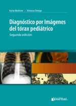
Diagnóstico por imagenes del tórax pediátrico – Moenne
$241,000Publicada con gran éxito en 2005, la primera edición de este libro se convirtió en una de las obras de referencia en español para interpretar los estudios de imágenes del tórax pediátrico.
Esta segunda edición fue revisada en su totalidad por las autoras, quienes agregaron nuevas imágenes de gran interés para ilustrar las patologías. El libro cuenta con capítulos sobre anatomía del sistema respiratorio pediátrico, técnicas de imágenes y protección radiológica, tórax normal, infecciones respiratorias bajas, alteraciones de la vía aérea y malformaciones congénitas, entre otros. Además de la actualización de los temas, se abordan también las nuevas modalidades diagnósticas.$283,000 -

Osborn’s Brain, 2nd Edition
$1,730,000Comprehensive, visually appealing, and easy to understand, Osborn’s Brain, second edition, by the highly esteemed Dr. Anne G. Osborn, provides a solid framework for understanding the complex subject of brain imaging when studied cover to cover. Almost completely rewritten and featuring 75% new illustrations, it combines essential anatomy with gross pathology and imaging, clearly demonstrating why and how diseases appear the way they do. The most immediate emergent diagnostic topics are followed by nonemergent pathologies, integrating the most relevant information from Dr. Osborn’s entire career of accumulated knowledge, experience, and interest in neuropathology, neurosurgery, and clinical neurosciences.
New to this Edition
-
- Includes new WHO classifications of brain tumors, new entities including IgG4-related disease and CLIPPERS, new and emerging infectious diseases, and updated insights into brain trauma and brain degeneration
- Expert Consult™ eBook version included with purchase. This enhanced eBook experience allows you to search all of the text, figures, Q&As, and references from the book on a variety of devices.
Key Features
-
- Covers the «must-know» aspects of brain imaging together with spectacular pathology examples, relevant anatomy, and up-to-date techniquesin neuroradiology-perfect for radiologists, neuroradiologists, neurosurgeons, and neurologists at all levels
- Begins with emergent topics such as trauma, nontraumatic hemorrhage, stroke, and vascular lesions, followed by infections, demyelinating and inflammatory diseases, neoplasms, toxic-metabolic-degenerative disorders, and congenital brain malformations
- Features more than 4,000 stunning, high-resolution radiologic images and medical illustrations, all of which are annotated to describe the most clinically significant features
- Includes Dr. Osborn’s trademark summary boxes scattered throughout for quick review of essential facts, as well as the most recent and up-to-date references available
- Helps readers think clearly about diagnoses, types of diagnoses, and the various pathologies that can affect the brain
$1,831,000 -
-
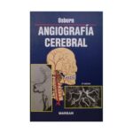
Angiografía Cerebral
Solicitar precioAl igual que sucede con todos mis libros, Angiografía Cerebral está concebido como un libro de enseñanza. He intentado unir, consolidar, simplificar e ilustrar la patología, epidemiología y, por supuesto, los hallazgos clínicos relativos a cada una de las principales enfermedades (y también de las menos importantes) que afectan a los vasos craneocerebrales. Para un repaso más fácil, los puntos clave se han resumido y destacado mediante cuadros que se incluyen en cada capítulo.
-
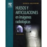
Huesos y Articulaciones en imágenes radiológicas
Solicitar precioLas primeras incursiones de Resnick en práctica de la radiología fueron la entrega de informes de su padre (también radiólogo especializado en la imagen musculoesquelética) a los médicos que habían ordenado estudios de rayos X, por lo general para confirmar fracturas sospechosas. Sus mas de cuarenta años de experiencia en el mundo de la radiología avalan este clásico que continúa siendo el libro de consulta mas completo del mercado.
En la actualidad los métodos diagnósticos aplicados de forma rutinaria en el análisis de los trastornos musculoesqueléticos, incluyen no solo las radiografías convencionales, sino TC, RM, ecografías, gammagrafías y artografías. Estas técnicas son prácticamente las que aportan los nuevos contenidos y actualizan la tercera edición de este Best Seller» que contiene 4.000 imágenes.
-
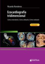
Ecocardiografía Tridimensional. Cómo entenderla, cómo utilizarla, cómo realizarla.
$519,000Las imágenes diagnósticas son un elemento esencial en la semiología cardiovascular en estos días. La tridimensionalidad es un aspecto indispensable para la visualización y comprensión de la anatomía y fisiopatología cardiovascular. En esta obra pionera en idioma español, el Dr. Ricardo Ronderos y sus colaboradores, todos ellos líderes internacionales en esta disciplina, dan cuenta del estado del arte en ecografía tridimensional y de sus perspectivas futuras. A lo largo de sus 32 capítulos, se desarrollan tanto los aspectos funcionales y normales como el diagnóstico de las principales patologías cardiacas mediante el uso de esta técnica. Ecografía tridimensional contiene un acceso a 197 videos en donde el autor explica todas las técnicas desarrolladas en el libro.
Una obra fundamental para aprender el uso de esta técnica transformadora del diagnóstico de la patología del corazón.$611,000


Be the first to review “RM Musculoesquelética”
You must be logged in to post a review.