
Eco de mama Handbook
Eco de mama Handbook – Lanfranchi
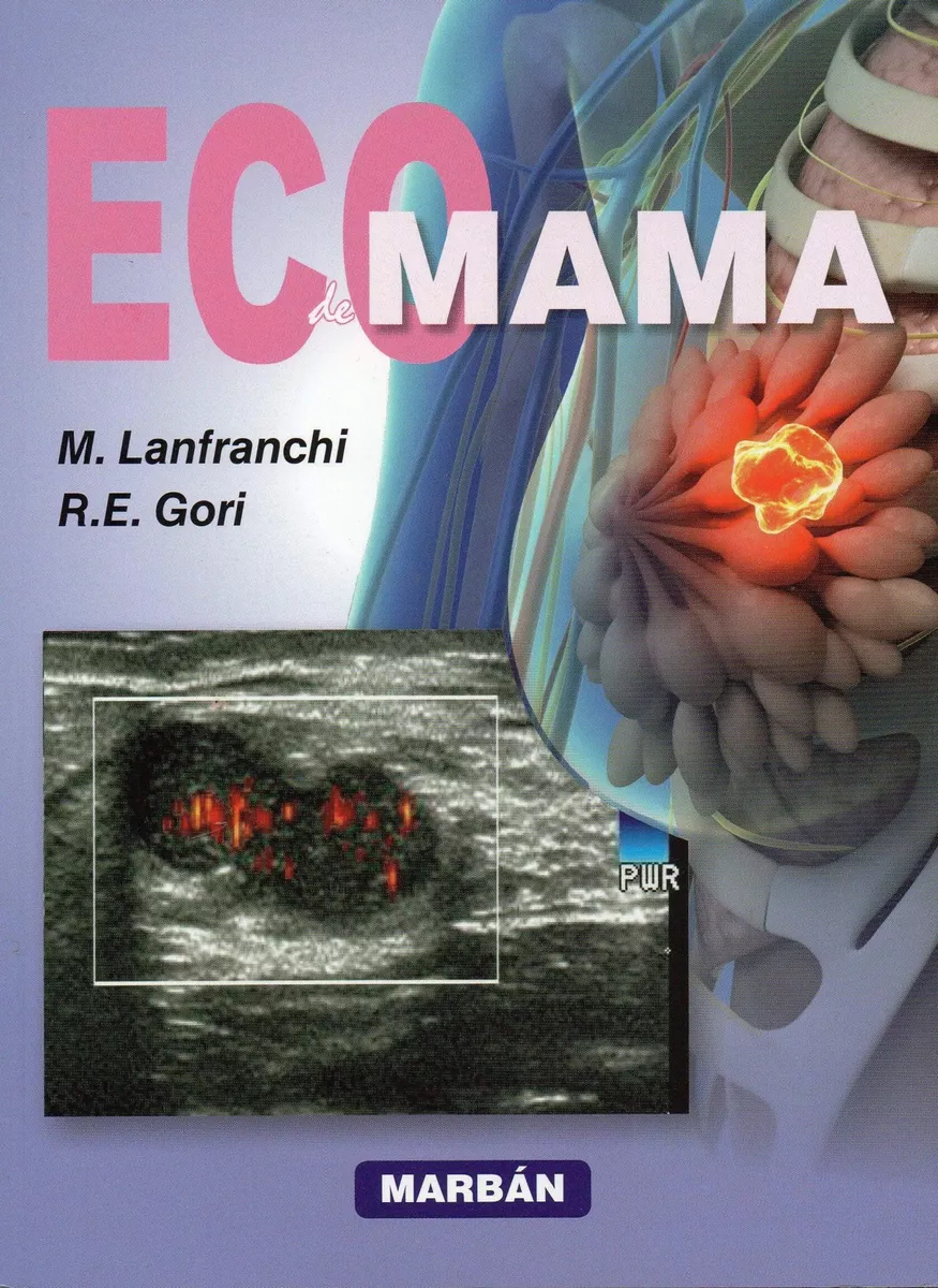
Este libro es un excelente tratado sobre la aplicación de los ultrasonidos en el diagnóstico de la patología mamaria. Proporciona una visión general de las técnicas actuales de ecografía y explica las ventajas e inconvenientes de cada una de ellas, ilustrando con imágenes de alta calidad las enfermedades malignas y benignas más importantes. Además, estas ilustraciones se enriquecen con flechas y etiquetas que aumentan su valor pedagógico y permitirán un diagnóstico más preciso y temprano.
Otra de las virtudes de este trabajo, es su absoluta “puesta al día” ya que refleja los procedimientos ecográficos de vanguardia y, para la organización y elaboración de los informes, se basa en los criterios BI-RAD correspondientes a la quinta edición publicada recientemente.
$155,000
Sin existencias
Related products
-
Avances en Diagnóstico por Imágenes: Cabeza y cuello
$245,000Los avances en el conocimiento de la patología de la cabeza y el cuello, región que integra la Neurorradiología junto con las patologías del sistema nervioso central y columna vertebral, nos motivaron a editar este nuevo volumen.
Con el fin de mantener la unidad editorial y la visión de conjunto, ofrecimos el cargo de Editor Invitado al Dr. Enrique Palacios Mayorga, médico neurorradiólogo de la Universidad de Tulane, a quien agradecemos haber aceptado hacerse cargo de esta tarea.
El contenido de Cabeza y Cuello se ha conformado con interesantes tópicos a cargo de autores cuya opinión es ampliamente reconocida por haberse destacado en su actividad profesional y académica en el campo de las neuroimágenes. Provenientes de diversos países (Canadá, Colombia, Estados Unidos, España, Panamá y México), estos profesionales actualizan el conocimiento y los avances sobre la anatomía de la cabeza y el cuello, la patología del plexo braquial, los senos paranasales y la órbita, y las neoplasias de cabeza y cuello, incluyendo laringe, adenopatías y base del cráneo. Asimismo, se incluyen conceptos diagnósticos sobre implantes dentales, sordera y alteraciones congénitas.
La importancia y actualidad de estos temas nos hacen confiar en que este noveno volumen, tal como los volúmenes precedentes de la Serie Avances en Diagnóstico por Imágenes, también será de utilidad en el desempeño profesional de los colegas y especialistas en formación. Por esta razón, agradecemos también al Dr. Palacios y a todos sus colaboradores por participar en este programa educativo que patrocina el Colegio Interamericano de Radiología.
-
Huesos y Articulaciones en imágenes radiológicas
Solicitar precioLas primeras incursiones de Resnick en práctica de la radiología fueron la entrega de informes de su padre (también radiólogo especializado en la imagen musculoesquelética) a los médicos que habían ordenado estudios de rayos X, por lo general para confirmar fracturas sospechosas. Sus mas de cuarenta años de experiencia en el mundo de la radiología avalan este clásico que continúa siendo el libro de consulta mas completo del mercado.
En la actualidad los métodos diagnósticos aplicados de forma rutinaria en el análisis de los trastornos musculoesqueléticos, incluyen no solo las radiografías convencionales, sino TC, RM, ecografías, gammagrafías y artografías. Estas técnicas son prácticamente las que aportan los nuevos contenidos y actualizan la tercera edición de este Best Seller» que contiene 4.000 imágenes.
-
MRI of the Prostate
$390,000Although prostate cancer is the second leading cause of cancer death in men in the USA, it can be treated successfully if detected early. Disease management has gradually changed to a paradigm that relies on close monitoring through active surveillance in select patients, as well as ongoing refinements in treatment interventions, including minimally invasive procedures. This has resulted in a critical need for a more exacting methodology for performing targeted biopsies, assessing risk levels, and devising treatment strategies. Prostate MRI has emerged as the most precise, state-of-the-art imaging modality for prostate cancer diagnosis and management, thereby creating an immediate demand for radiologists to become proficient in its use.
Conceived and edited by a leading authority, with contributions from renowned experts in the field, MRI of the Prostate: A Practical Approach is the first book to tackle this important topic. It provides an overview of the fundamentals of prostate MRI acquisition, interpretation, and reporting. Readers will benefit from a wide range of insightful perspectives gleaned from years of hands-on experience.
Key Highlights
- Prostate Imaging Reporting and Data System (PI-RADS) for prostate MRI interpretation and cancer risk scoring
- Clinical pearls on the optimization and application of prostate MRI for risk assessment, disease staging, MRI-targeted biopsy, recurrent disease, and active surveillance
- The emerging utilization of PET and PET/MRI for primary prostate cancer evaluation
- More than 700 illustrations with one entirely image-based chapter featuring educational case studies
Radiologists will learn how to optimally perform and interpret prostate MRI, and referring physicians will learn to integrate it into day-to-day practice. This book is an essential resource for radiologists and radiology residents, as well as urologists, oncologists, MRI technicians, and other medical practitioners who treat patients with genitourinary disorders.
-
Ecografía de testículo, bolsa escrotal y pene – Lanfranchi
$484,000El advenimiento de técnicas de alta resolución en diagnóstico por ultrasonidos permitió el desarrollo de la investigación de órganos y patologías inaccesibles en el pasado cercano. Las llamadas small parts o pequeñas partes comienzan a tomar un gran auge por los excelentes resultados clínicos y patológicos no solamente en el diagnóstico morfológico, sino también en el fisiológico -con el advenimiento del Doppler- y en la precisión de las punciones guiadas por ultrasonidos.
La profesora Dra. Mirta Ester Lanfranchi se identifica tempranamente con este nuevo y fascinante campo de la ecografía y desarrolla una intensa actividad asistencial, de investigación y docencia que supera largamente las fronteras de nuestro país. Preceden a esta obra los libros Ecografía mamaria, edición en español y en inglés; Ecografía tiroidea y, ahora, Ecografia de testículo, bolsa escrotal y pene.
Existe escasa bibliografía que concentre esta temática en un libro de fácil y amena lectura.
El desarrollo de los temas está precedido por un conciso texto relativo a la embriología, la semiología clínica y la historia clínica de cada una de las enfermedades tratadas.
La lectura de esta obra puede encararse como la de un preciso libro de texto o como la de un atlas con profusión de imágenes, con claros epígrafes descriptivos de los principales signos presentes en el estudio ecográfico. La facilidad de palabra de la Dra. Lanfranchi fluye en este texto con la frescura y facilidad con que se escuchan sus conferencias en vivo.
Auguro un éxito trascendente de este libro no solamente en el área del Diagnóstico por Imágenes, sino también por la aplicación de estos precisos resultados en la práctica de las diferentes especialidades intervinientes: urólogos, cirujanos, pediatras, andrólogos, sexólogos y médicos en general que vivieron las limitaciones de explorar estos órganos sin estos fantásticos avances.Del Prólogo del Prof. Dr. Carlos A Bruguera
$569,000


Be the first to review “Eco de mama Handbook – Lanfranchi”
You must be logged in to post a review.