
-
Ecografía en Dermatología y Dermoestética 2ed. – Alfageme
$253,000En los últimos años, la aplicación de los ultrasonidos al diagnóstico de las enfermedades y alteraciones estéticas cutáneas ha experimentado un desarrollo exponencial. La segunda edición de este manual, imprescindible referencia para los médicos que realizan exploraciones en el campo de la ecografía, facilita al lector las ultimas actualizaciones de esta técnica de forma clara, sencilla y completa.
-
Diseases of the Abdomen and Pelvis 2023-2026 – Hodler
$290,000This open access book deals with imaging of the abdomen and pelvis, an area that has seen considerable advances over the past several years, driven by clinical as well as technological developments. The respective chapters, written by internationally respected experts in their fields, focus on imaging diagnosis and interventional therapies in abdominal and pelvic disease; they cover all relevant imaging modalities, including magnetic resonance imaging, computed tomography, ultrasound, and positron emission tomography. As such, the book offers a comprehensive review of the state of the art in imaging of the abdomen and pelvis.
IDKD books are extensively re-written every four years. As a result, they offer a comprehensive review of the state of the art in imaging. The book is clearly structured with learning objectives, abstracts, subheadings, tables and take-home points, supported by design elements to help readers easily navigate through the text. As an IDKD book, it is particularly valuable for general radiologists, radiology residents, and interventional radiologists who want to update their diagnostic knowledge, and for clinicians interested in imaging as it relates to their speciality.
-
Pediatric Imaging A Core Review – Blumer
$332,000Prepare for success on the pediatric imaging component of the radiology Core Exam! Pediatric Imaging: A Core Review, 2nd Edition, by Drs. Steven L. Blumer, David M. Biko, and Safwan S. Halabi, is an up-to-date, practical review tool written specifically for the Core Exam. This helpful resource contains over 300 image-rich, multiple-choice questions with detailed explanations of right and wrong answers, revised content, and additional eBook questions to ensure you’re ready for the Core Exam or recertification exam.
$368,000 -
Neuroradiology A Core Review 2ed. – Dubey
$345,000Prepare for success on the neuroimaging component of the radiology Core Exam! Neuroradiology: A Core Review, 2nd Edition, by Drs. Sathish Kumar Dundamadappa, Prachi Dubey, Daniel Thomas Ginat, and Gul Moonis, is an up-to-date, practical review tool written specifically for the Core Exam. This helpful resource contains 500 image-rich, multiple-choice questions with detailed explanations and annotated images of right and wrong answers, fully revised content, high-yield tables for easy review, and additional eBook questions to ensure you’re ready for the Core Exam or recertification exam.
-

Diagnostic Ultrasound: Abdomen and Pelvis 2nd Edition – Kamaya
$1,422,000Develop a solid understanding of ultrasound of the abdomen and pelvis with this practical, point-of-care reference in the popular Diagnostic Ultrasound series. Written by leading experts in the field, the second edition of Diagnostic Ultrasound: Abdomen and Pelvis offers detailed, clinically oriented coverage of ultrasound imaging of this complex area and includes illustrated and written correlation between ultrasound findings and other modalities. The most comprehensive reference in its field, this image-rich resource helps you achieve an accurate ultrasound diagnosis for every patient.
$1,580,000 -
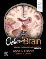
Osborn’s Brain 3rd Edition
$1,545,000Combining informative, meticulously crafted prose with more than 4,000 high-quality images, Osborn’s Brain, third edition, is a comprehensive, easy to understand, and visually stunning learning curriculum from highly esteemed author Dr. Anne G. Osborn. This fully revised edition provides a solid framework for understanding the complex subject of brain imaging, integrating relevant information from Dr. Osborn’s entire career of accumulated knowledge, experience, and interest in neuropathology, neurosurgery, and clinical neurosciences. While neuroradiologists will find intriguing, thought-provoking insights included especially for them in every chapter, Osborn’s Brain is an excellent review resource for physicians at all levels of expertise—from seasoned radiologists and neurosurgeons to new and senior residents or fellows.
-
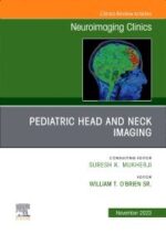
Pediatric Head and Neck Imaging, An Issue of Neuroimaging Clinics – O’Brien
$455,000In this issue of Neuroimaging Clinics, guest editor Dr. William T. O’Brien, Sr., brings his considerable expertise to the topic of Pediatric Head and Neck Imaging. The differential diagnosis of neck masses in pediatric patients differs compared to masses that arise in adults. In this issue, top experts in the field discuss imaging of hearing loss, sinus infections, neck masses, and congenital lesions, and more.Key Features- Contains 11 relevant, practice-oriented topics including conductive hearing loss in children; congenital cystic neck masses; common solid and vascular neck masses in children; pediatric facial and paranasal sinus infections; infectious and inflammatory processes of the orbits in children; and more.
- Provides in-depth clinical reviews on pediatric head and neck imaging, offering actionable insights for clinical practice.
- Presents the latest information on this timely, focused topic under the leadership of experienced editors in the field. Authors synthesize and distill the latest research and practice guidelines to create clinically significant, topic-based reviews.
-
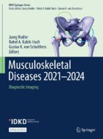
Musculoskeletal Diseases 2021-2024 – Hodler
$290,000This open access book focuses on imaging of the musculoskeletal diseases. Over the last few years, there have been considerable advances in this area, driven by clinical as well as technological developments. The authors are all internationally renowned experts in their field. They are also excellent teachers, and provide didactically outstanding chapters. The book is disease-oriented and covers all relevant imaging modalities, with particular emphasis on magnetic resonance imaging. Important aspects of pediatric imaging are also included.
IDKD books are completely re-written every four years. As a result, they offer a comprehensive review of the state of the art in imaging. The book is clearly structured with learning objectives, abstracts, subheadings, tables and take-home points, supported by design elements to help readers easily navigate through the text. As an IDKD book, it is particularly valuable for general radiologists, radiology residents, and interventional radiologists who want to update their diagnostic knowledge, and for clinicians interested in imaging as it relates to their specialty.
-
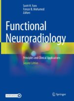
Functional Neuroradiology Principles and Clinical Applications – Faro
$1,190,000This new edition fully updates and expands Faro and Mohamed’s Functional Neuroradiology, a gold standard, comprehensive introduction to the state-of-the-art functional imaging in neuroradiology, including the physical principles and clinical applications of Diffusion, Perfusion, Permeability, MR spectroscopy, Positron Emission Tomography, BOLD fMRI and Diffusion Tensor Imaging.
With chapters written by internationally distinguished neuroradiologists, neurologists, psychiatrists, cognitive neuroscientists, and physicists, Functional Neuroradiology is divided into 12 major sections, including: Diffusion and Perfusion Imaging, Magnetic Resonance Spectroscopy and Chemical Exchange Saturation Transfer Imaging, Multi-Modality Functional Neuroradiology, BOLD Functional MRI, Diffusion Tensor Imaging, Presurgical Brain Tumor Mapping, Emerging neuroimaging techniques, Functional Spine and Hydrocephalus imaging, and Neuroanatomical Gray and White matter Brain Atlases. This second edition is fully updated throughout and includes more than 15 new chapters on topics such as: Brain tumor Radiogenomics, CNS Tumor Surveillance and Functional MR Perfusion Imaging, CNS Machine Learning, Focused Ultrasound therapy, TBI Sports Related Injury, and CNS Lymphatic system.
By offering readers a complete overview of functional imaging modalities and techniques currently used in patient diagnosis and management, as well as emerging technology, Functional Neuroradiology is a vital information source for physicians and cognitive neuroscientists involved in daily practice and research.
-
Dermatologic Ultrasound with Clinical and Histologic Correlations Softcover – Wortsman
$798,000Significant technological advances have produced equipment that allows imaging of the skin with variable frequency ultrasound in previously unseen detail and provides a range of dynamic data that is currently unmatched by any other technology. Dermatologic Ultrasound with Clinical and Histologic Correlations is a comprehensive introduction to ultrasonography of the skin, nails, and scalp as it relates to the assessment and diagnosis of dermatologic diseases. It provides radiologists, sonographers, dermatologists, and physicians with interest in skin imaging with a concise understanding of the diagnosis of dermatologic conditions through extensive high-resolution gray scale and color Doppler ultrasound images and presents classical correlations of clinical dermatologic lesions with sonographic and histologic findings. Featuring more than 1700 images, this text-atlas provides an excellent starting point in learning about this topic.
Featuring contributions from world-renowned authorities in the field of superficial ultrasound imaging, the book reviews the technical considerations relating to color Doppler ultrasound of the skin; surveys the dermatologic entities that can be visualized with ultrasound imaging, such as cutaneous tumors, inflammatory diseases, hemangiomas and vascular malformations, melanoma, nail tumors, scalp diseases and cosmetic conditions; shows common simulators of cutaneous diseases; and discusses protocols for assessing common dermatologic conditions. Inclusion of clinical overviews, tips, and pitfalls enables a better understanding of the pathologies of the disorders and the methodological approach in assessing these entities.
-
Xu / Diagnostic Ultrasound in Dermatology
$663,000This book offers readers details in application of high-frequency ultrasound in dermatology, a new method playing increasingly important roles in diagnosis of skin diseases. At first, chapters introduce anatomy and ultrasound features of normal skin. Then terminology, image quality, and artifact of dermatologic ultrasound are presented. After that, ultrasound features of benign and malignant skin tumors, inflammation, autoimmune disease, and traumas are described with diagnostic tips for specific disease. It will be a valuable reference book not only for dermatologist and radiologist, but also for plastic surgeon and cosmetologist.
$698,000 -
Kandarpa Handbook of Interventional Radiology 2023 6ed.
$378,000Focusing on time-tested protocols, tailored imaging and current procedural equipment, this popular, practical handbook by Drs. Krishna Kandarpa, Lindsay Machan, Robert Lewandowski, and Parag J. Patel features extensive updates to keep you current with rapid growth in the field. Now in brilliant full color throughout, Kandarpa Handbook of Interventional Radiologic Procedures, 6th Edition, is a convenient, easy-access guide to all current radiologic procedures. It’s an ideal resource not only for practicing interventional and general radiologists, but also for fellows and residents in training, IR nurses, and special procedure technologists.
-
Breast Imaging A Core Review 3ed – Shah
$345,000Prepare for success on the breast imaging component of the radiology Core Exam! Breast Imaging: A Core Review, 3rd Edition, by Drs. Biren A. Shah and Sabala Mandava, is an up-to-date, practical review tool written specifically for the Core Exam. This helpful resource contains 300 image-rich, multiple-choice questions with detailed explanations of right and wrong answers, fully revised content, high-yield tables for easy review, and additional eBook questions to ensure you’re ready for the Core Exam or recertification exam.
-
Medicina nuclear. Fundamentos – Jadvar
$397,000La medicina nuclear está evolucionando como uno de los campos más prometedores de la medicina clínica. La reciente aceleración en el desarrollo y la aprobación de radiofármacos para la obtención de imágenes y la terapia dirigida ha contribuido de forma significativa a la atención de pacientes con diversas enfermedades en los ámbitos de la cardiología, la neurología, la oncología y las enfermedades inflamatorias e infecciosas.
Medicina nuclear. Fundamentos presenta una visión completa y concisa de los conocimientos más importantes de este desafiante campo en evolución. Es una obra completa para que residentes e internos la utilicen durante las rotaciones, o como un repaso rápido para radiólogos y médicos de medicina nuclear en activo.
Se abarcan todos los temas clínicos principales, incluyendo información adicional sobre los fundamentos de la física de la medicina nuclear, tecnología de los equipos y garantía de calidad, radioquímica, y seguridad de la radiación, además de contenidos sobre temas especializados como el embarazo, la lactancia, la pediatría y la infección por SARS-CoV-2 (covid-19).$467,000 -
Fundamentals of Pediatric Imaging 3ed. – Donnelly
$450,000Fundamentals of Pediatric Imaging, Third Edition presents the foremost techniques of pediatric medical image analysis and processing. It includes advanced imaging techniques, neuro applications, and highlights basic anatomy needed to understand this complex specialty. The book introduces the theory and concepts of pediatric digital image analysis and newly revised information on quality and safety topics, imaging modalities, imaging applications, and new discoveries in diseases and treatments. The newly revised edition provides updates in areas of expertise including neurologic, musculoskeletal, cardiac, chest, and GU imaging. Edited by Lane F. Donnelly, MD, recipient of the Society of Pediatric Radiology’s 2009 Singleton-Taybi Award, this book is sure to be a prime reference in pediatric medical imagining.
$499,000 -
MRI and Traumatic Brain Injury, An Issue of Neuroimaging Clinics of North America – Maralani
$480,000In this issue of Neuroimaging Clinics, guest editors Drs. Pejman Jabehdar Maralani and Sean Symons bring their considerable expertise to the topic of Neurotrauma. Top experts in the field cover key topics such as conventional MRI in trauma management in adults and children; imaging approach to concussion; clinical updates on concussion; the current state of DWI/DTI for trauma prognostication; the current state of fMRI/rs-fMRI for trauma prognostication; and more.
-

Physical Medicine and Rehabilitation Secrets – O’Young
$330,000For more than 30 years, the highly regarded Secrets Series® has provided students, academics, and practitioners in all areas of health care with concise, focused, and engaging resources for quick reference and exam review. Physical Medicine and Rehabilitation Secrets, 4th Edition, offers practical, up-to-date coverage of the full range of essential topics in this dynamic field. This highly regarded resource features the Secrets’ popular question-and-answer format that also includes lists, tables, weblinks, pearls, memory aids, and an easy-to-read style – making an inquiry, reference, and review quick, easy, and enjoyable.
Key Features
- The proven Secrets Series® format gives you the most return for your time – concise, easy to read, engaging, and highly effective.
- Fully revised and updated, including new information on geriatric rehabilitation, rehabilitation philosophy, vocational rehabilitation, disability rating and impairments, and legislation and reimbursement.
- New chapters and content include Longitudinal Learning; Regenerative Medicine; Musculoskeletal Ultrasound, PM&R ideology and Disability Awareness & Sensitivity, Organ Transplantation; Spinal Deformity: and more.
- Top 100 Secrets and Key Points boxes provide a rapid overview of the secrets you must know for success in practice, exams, and teaching sessions.
- Bulleted lists, mnemonics, and practical tips from global leaders in the field provide a concise overview of important board-relevant content.
- Portable size makes it easy to carry with you for quick reference or review anywhere, anytime.
- Enhanced eBook version included with purchase. Your enhanced eBook allows you to access all of the text, figures, and references from the book on a variety of devices.
-
ExpertDDx: Brain and Spine – Salzman
$1,707,000Designed with the busy practitioner in mind, ExpertDDx: Brain and Spine, third edition, quickly guides you to the most likely differential diagnoses based on key imaging findings and clinical information. This superbly illustrated resource covers more than 275 of the top differential diagnoses across a broad spectrum of central nervous system diseases, presenting parallel groups of anatomically based differentials, generic imaging patterns, modality-specific findings, and clinically based differentials for each area. Now fully revised and up-to-date, this practical reference clearly guides you through useful, actionable differential diagnoses that lead to definitive findings in every area of the brain and spine.Key Features- Presents multiple clear, sharp, succinctly annotated images for each diagnosis; a list of diagnostic possibilities sorted as common, less common, and rare but significant; and brief, bulleted text offering helpful diagnostic clues
- Reflects changes in 2021 WHO CNS tumor grading and nomenclature
- Contains newly identified entities, new differential diagnoses, and updated references
- Shows both typical and variant manifestations of each possible diagnosis
- Includes more than 7,000 high-quality print and online images
- Features updated genetic information now available in Online Mendelian Inheritance in Man (OMIM)
- Separates adult and pediatric DDx lists for even faster reference
- Assists you in building either a definitive diagnosis from an imaging study or a carefully refined list of reasonable differential diagnoses
- Includes an eBook version that enables you to access all text, figures, and references, with the ability to search, customize your content, make notes and highlights, and have content read aloud
-
Core Radiology A Visual Approach to Diagnostic Imaging – Sun
$890,000Embodying the principle of ‘everything you need but still easy to read’, this fully updated edition of Core Radiology is an indispensable aid for learning the fundamentals of radiology and preparing for the American Board of Radiology Core exam. Containing over 2,100 clinical radiological images with full explanatory captions and color-coded annotations, streamlined formatting ensures readers can follow discussion points effortlessly. Bullet pointed text concentrates on essential concepts, with text boxes, tables and over 400 color illustrations supporting readers’ understanding of complex anatomic topics. Real-world examples are presented for the readers, encompassing the vast majority of entitles likely encountered in board exams and clinical practice. Divided into two volumes, this edition is more manageable whilst remaining comprehensive in its coverage of topics, including expanded pediatric cardiac surgery descriptions, updated brain tumor classifications, and non-invasive vascular imaging. Highly accessible and informative, this is the go-to introductory textbook for radiology residents worldwide.
-
Tomografía Computarizada Cardíaca Principios, técnica y aplicaciones clínicas – Bastarrika
$234,000La tomografía computarizada (TC) cardíaca se ha convertido en la técnica de elección para la valoración no invasiva de la vascularización coronaria. Su utilidad se basa en la capacidad de esta técnica para descartar la enfermedad coronaria, pero su potencial trasciende a la mera valoración de la permeabilidad de la luz del vaso, ya que también permite caracterizar la pared vascular y establecer la composición de la placa de ateroma.
Tomografía computarizada cardíaca. Principios, técnica y aplicaciones clínicas trata los fundamentos de la TC cardíaca desde sus principios esenciales para adentrarse en los aspectos más avanzados de la técnica. Esta obra se caracteriza por:
- Estar escrita con un estilo práctico, tal y como lo demuestran los algoritmos, tablas y figuras que comprende cada capítulo.
- Contar con trucos y consejos para que el profesional adquiera y repase los conocimientos necesarios para realizar e interpretar estudios cardíacos por TC.
- Permitir al lector conocer las bases teóricas de la TC cardíaca, comprender los protocolos de adquisición, interpretar las exploraciones y familiarizarse con las aplicaciones clínicas establecidas y en investigación.
La obra está dirigida a los profesionales de la salud interesados en la imagen cardíaca, especialmente a radiólogos y cardiólogos. También resulta de interés para técnicos, enfermeras de radiología y estudiantes de esta materia.
$260,000 -
Central Nervous System Infections, An Issue of Neuroimaging Clinics
$480,000Central Nervous System Infections
In this issue of Neuroimaging Clinics, guest editor Dr. Tchoyoson Lim Choie Cheio brings his considerable expertise to the topic of Central Nervous System Infections. Infections can involve any part of the CNS and often, multiple parts of the CNS are involved at the same time. The imaging surrounding them is constantly evolving, and in this issue, key international experts provide a thorough update of the imaging of these common and pervasive infections.Key Features- Contains 13 practice-oriented topics including emerging public health, multidisciplinary teams and pitfalls in CNS infection imaging; acute neurological complications of COVID-19; subacute to chronic neuroimaging findings in SARS-CoV-2/COVID-19 infection; imaging of opportunistic infections and HIV/AIDS; imaging of head and neck infections; and more.
- Provides in-depth clinical reviews on central nervous system infections, offering actionable insights for clinical practice.
- Presents the latest information on this timely, focused topic under the leadership of experienced editors in the field. Authors synthesize and distill the latest research and practice guidelines to create clinically significant, topic-based reviews.
-
Interventional Ultrasound of the Breast – Fornage
$736,000This book is the premier guide to ultrasound-guided interventions of the breast. Written by Bruno D Fornage, a world-renowned leader in the fields of breast ultrasound and ultrasound-guided interventions, it covers in detail techniques of freehand ultrasound-guided breast biopsy, placement of post-biopsy markers, localization of nonpalpable lesions, and percutaneous ablation of breast masses. A large part of the book describes the highly effective combined use of fine-needle aspiration and core-needle biopsy during the staging of breast cancer; this combination has been used successfully by the author at MD Anderson Cancer Center for 3 decades.
Throughout the book, the author shares numerous tips and tricks, many of which have not been published before. With over 1300 figures and and 200 videoclips depicting ultrasound-guided procedures, Interventional Ultrasound of the Breast is the authoritative resource for breast imagers, interventional radiologists, surgical breast oncologists, pathologists, and anyone who embarks on ultrasound-guided breast biopsies and other breast procedures. In addition, the techniques described in this book are applicable to many other areas of the body, including the thyroid and soft tissues.
$820,000 -
Search Pattern: A Systematic Approach to Diagnostic Imaging – Long Tu
$176,000Search Pattern is a collection of step-by-step guides to more than a hundred of the most common types of studies in radiology. Blind spots reported in the literature as well as practical wisdom from experts is synthesized into highly structured processes that can guide the development of better practice. Much of the contained insight has never been organized in one place before. Search Pattern covers almost every type of study that a radiologist will encounter in training or practice. This text is written with the assumption that the reader has familiarity with basic radiologic terminology, anatomy, and physics. In the interest of brevity, almost all information outside of the organized approaches is omitted. The reader is encouraged to look up terms, images, and background information from supplementary resources. Formalized teaching of search patterns is a missing part of the educational literature in our field. Hopefully this book helps fill that void. It is one that I would have benefited from greatly when I was a resident. – Long Tu, YDR Class of 2020
-
Textbook of Dermatologic Ultrasound – Wortsman
$850,000This book provides a pedagogical guide to dermatologic ultrasound. As in any imaging field, dermatologic ultrasound is dynamic, and the area expands with the release of new technology. This textbook is a mix of the essential knowledge necessary to start and a detailed update to the dermatologic ultrasound field.
This book is divided into three sections: The Requisites, Ultrasound Features of Common Dermatologic Conditions, and Practicalities. The first section details the basic information needed in dermatologic ultrasound, including technical recommendations and dermatologic concepts. The second covers major dermatologic conditions and their ultrasound presentation, including benign and malignant skin tumors, pediatric conditions, nail entities, inflammatory conditions, infections and infestations, and aesthetics. The final section covers tips for reporting and interventional dermatologic ultrasound procedures.Chapters present substantial clinical, ultrasonographic, and histologic correlation, including algorithms to help discriminate different conditions. The book additionally includes 150 self-assessment questions (CME) in the backmatter, multiple key points, a plethora of state-of-the-art images with probes that go from 18 to 70 MHz, and ultrasound videos.
This is a must guide for physicians, residents, and students in radiology, dermatology, plastic surgery, and any professional who wants to brush up on the dermatologic ultrasound field.
-
Pie y Tobillo – Quesada
$218,000Pie y Tobillo
El trabajo del especialista en una unidad de Cirugía Ortopédica y Traumatología es una tarea compleja que requiere el conocimiento de numerosas patologías. Esta obra pretende ser una herramienta de consulta útil para los residentes en la especialidad que resuelva los problemas más habituales a los que deben enfrentarse en su primera etapa formativa en el hospital.
$257,000


