
- No puedes añadir «Search Pattern: A Systematic Approach to Diagnostic Imaging - Long Tu» al carrito porque el producto está agotado.
-
Clark’s Essential Guide to Clinical Ultrasound – Dodgeon
$176,000This easy-to-understand pocketbook in the highly respected Clark’s stable of diagnostic imaging texts is an invaluable tool for students, sonographers and other ultrasound practitioners, providing practical guidance on clinical ultrasound procedures, summarising current relevant literature and professional body guidelines.
The content is arranged by anatomical system and organ for ease of reference, with each section comprising a short introduction, the indications for the procedure, patient preparation, the imaging procedure itself, along with an image analysis, supported throughout by positioning photographs and clinical images.
Clark’s Essential Guide to Clinical Ultrasound is ideal for all users of clinical ultrasound at both undergraduate and postgraduate level and will also provide a convenient distillation of the latest best practice and guidelines for sonographers, midwives, vascular technologists, ECG technologists, medical doctors, sports injury specialists, paramedics, and other health professionals.
-
Ultrasound of Congenital Fetal Anomalies – Paladini
$1,247,000An acclaimed overview of ultrasound for the prenatal diagnosis of congenital anomalies returns in a new enlarged edition. In particular, the coverage of both Central Nervous System congenital and acquired anomalies as well as Congenital Heart Disease has been expanded enormously, to make this an impressive comprehensive resource for Fetal Neurology and Fetal Cardiology. Together with additional new chapters on guidelines and protocols, equipment, and disorders of sexual differentiation, and new insight into fetal surgery procedures, this third edition almost becomes three books in one.
-
Diagnostic Imaging: Cardiovascular 3rd Edition – Abbara
$1,445,000Covering the entire spectrum of this fast-changing field, the third edition of Diagnostic Imaging: Cardiovascular, Third Edition is an invaluable, everyday resource for general, cardiac, thoracic, and vascular radiologists as well as cardiologists and trainees—anyone who requires an easily accessible, highly visual reference in this complex area of imaging. Drs. Suhny Abbara, Seth Kligerman, and a team of highly regarded experts provide up-to-date information in concise yet detailed chapters to help you make informed decisions at the point of care. The text is meticulously illustrated, precisely delineated, and extensively referenced, making it a useful learning tool for readers at all levels of experience as well as a handy reference for daily clinical practice.Key Features- Provides comprehensive, expert information on all aspects of cardiac and vascular imaging to help you quickly determine the correct diagnosis for patients with symptoms or indications of cardiovascular disease
- Contains 200+ fully updated chapters on specific areas or groups of related diseases, with in-depth information ranging from common diagnoses to the rare and obscure; each topic is supported by multimodality imaging examples with detailed discussions from experts in the field
- Features new chapters on the expanded use of AI in cardiovascular imaging, as well as multimodality advances in PET and MR for inflammatory cardiomyopathies and in CT and MR for advances in myocardial blood flow imaging
- Discusses new imaging guidelines for the management of acute and chronic chest pain and new transcatheter procedures
- Covers the latest CAD-RADS 2.0 classification, COVID-related vascular complications, and novel dose-reduction techniques
- Includes access to new videos that promote a visual understanding of cardiac flow and normal vs. abnormal cardiovascular health, among other topics
- Reflects new material throughout based on WHO updates for tumors and pulmonary hypertension
- Features more than 2,300 high-quality print images (with an additional 1,300+ images in the complimentary eBook), including videos, radiologic images, full-color illustrations and graphics, histologic images, and clinical photographs
- Uses a bulleted, image-rich layout with short chapters designed for quick access to critical content on terminology, characteristics seen in various imaging studies on the same anatomic location, differential diagnoses, pathologic and clinical issues to consider, and more
- Includes an eBook that enables you to access everything in this print version as well as additional text, images, and videos with the ability to search, customize your content, make notes and highlights, and have content read aloud
-
Specialty Imaging: HRCT of the Lung, 3rd Edition – Martinez
Solicitar precioPart of the highly regarded Specialty Imaging series, HRCT of the Lung, third edition, reflects the many recent changes in HRCT diagnostic interpretation. An easy-to-read bulleted format and thousands of state-of-the-art imaging examples guide you step by step through every aspect of thin-section CT and HRCT in the evaluation of patients with suspected lung disease. This book is an ideal resource for radiologists and internal medicine specialists who need an easily accessible tool to help them understand the indications, strengths, and limitations of HRCT in their practice.Key Features- Helps you identify and characterize critical HRCT findings and gain a solid understanding of cross-sectional imaging anatomy of the lung—crucial skills for formulating diagnoses and appropriate differential diagnoses
- Delivers details on anatomy-based exploration as well as nodules and micronodules, cysts and pseudocysts, reticulation and honeycombing, mosaic attenuation, ground-glass opacities, and interlobular septal thickening
- Uses a time-saving, bulleted format that distills essential information for fast and easy comprehension
- Includes new chapters on post-COVID-19 findings, respiratory syncytial virus, and e-cigarette and vaping product use-associated lung injury (EVALI), as well as content on new morphologic patterns for pulmonary fibrosis, the new classification for hypersensitivity pneumonia, and more
- Provides direction for determining common, less common, and rare diagnoses based on morphologic features, distribution of abnormalities, and/or histologic patterns
- Features superb illustrations with comprehensive captions that display both typical and variant findings on HRCT scans
- Offers new references, new images, and new histopathologic correlations throughout
- Includes an eBook version that allows you to access everything in this print version as well as additional images, text, and references, with the ability to search, customize your content, make notes and highlights, and have content read aloud; additional digital ancillary content may publish up to 6 weeks following the publication date
-
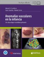
Anomalías Vasculares en la Infancia – Cordisco
$517,000Anomalías vasculares en la infancia | Un abordaje multidisciplinario es el resultado del trabajo interdisciplinario entre especialistas de dermatología, radiología, genética, cirugía, hematología y patología.
Los más recientes hallazgos genéticos han abierto un nuevo horizonte para la atención de estas complejas entidades con la utilización de novedosas terapias dirigidas, lo que permite obtener mejores resultados y ofrecer a los pacientes una mejor calidad de vida.
Principales características:
– Escrito por referentes mundiales, la obra aporta información actualizada y relevante, presentada de manera clara y ordenada, acerca de los últimos avances en estas complejas entidades.
– Contenido estructurado en 22 capítulos, que despliegan exhaustivamente el rasgo interdisciplinario de la obra.
– Aporta una gran cantidad de fotografías de importante valor científico para el estudio, diagnóstico y detección de estas enfermedades raras y poco frecuentes.
– Incorpora la clasificación de las anomalías vasculares actualizada en el último congreso de la Sociedad Internacional para el Estudio de las Anomalías Vasculares [ISSVA], realizado en Madrid en mayo de 2024.Anomalías vasculares en la infancia | Un abordaje multidisciplinario está dirigido a dermatólogos, pediatras, radiólogos, cirujanos, patólogos, hematólogos, genetistas y a los especialistas de otras áreas interesados en el manejo interdisciplinario de estas enigmáticas patologías. Su aporte también es relevante para investigadores académicos en el campo de la medicina vascular.
$608,000 -
Fetal Neurosonography – Volpe
$675,000What would make this book unique is its focus on the fetal central nervous system (CNS) ultrasound, encompassing the wide range of CNS anomalies and the need for in-depth knowledge in fetal brain and spine normal anatomy and related abnormalities. From the anatomy and pathophysiology of the main CNS anomalies to the latest advancements in diagnostic techniques and therapeutic interventions, this book provides a thorough exploration of the subject matter.
One of the key strengths of this book series lies in its structured approach. Each volume follows an organized framework, enabling readers to navigate through the content effortlessly. From the fundamentals of CNS ultrasound imaging to the intricacies of differential diagnosis, counseling and management of CNS anomalies, this book presents information in a logical and accessible manner. This structure not only facilitates comprehension but also allows readers to quickly locate specific topics and delve deeper into areas of interest.
By creating this book, our aim is to fill a critical gap in the field of fetal medicine and ultrasound education. While numerous resources exist, this book stands out due to its comprehensive nature, covering both the theoretical foundations and practical applications of neurosonography. It also offers essential information for counseling and presents high-quality ultrasound images, enabling specialists to systematically and confidently approach normal CNS anatomy and accurately diagnose and manage CNS abnormalities, also exploring the role of additional imaging techniques, such as MRI, and genetic testing.
This book bridges the divide between research and clinical practice, equipping healthcare professionals with the necessary knowledge and skills to provide optimal care for women throughout their reproductive lifespan.
In conclusion, this book offers a resource for healthcare professionals, researchers, educators, and students in the field, bridging the gap between theory and practice and aiming to elevate the standard of care in fetal neurosonography.
-
EDiR – The Essential Guide – Oleaga
$499,000The main objective of this new edition is to facilitate the learning of radiology through a handbook that includes all disciplines of radiology. It is aimed to guide young trainees from the beginning of their radiology training and help them in the preparation of the European Diploma in Radiology (EDiR).
The 2nd edition features new cases in all the subspecialities, now including also Hybrid Imaging, Informatics (with some information regarding Artificial Intelligence) and Physics.
The book is intended to be a reference in the field of radiology providing a standardised study opportunity of the specialty following the outline of EDiR. The content has been selected and supervised by the members of the different committees responsible for the examination.
Each chapter is structured in three sections similar to the European exam: a first section with Multiple Response Questions (MRQs), a second section with Short Cases (SCs) and, finally, cases of clinicalreasoning (CORE). For a deeper understanding, this new edition features 30 exclusive videos that complement the text. These engaging visuals enhance the learning experience, making complex concepts easier to grasp.
The reader is exposed to questions and problems as they arise in the European examination. It covers technical, security and management aspects, all of which are included in the European curriculum and which form part of the content of the exam.
-
Diseases of the Chest, Heart and Vascular System 2025-2028 – Hodler
$250,000This open access book offers an essential overview of chest, heart and vascular system imaging. Over the last few years, there have been considerable advances in this area, driven by both clinical and technological developments. Written by leading international experts and teachers, the chapters are disease-oriented and cover all relevant imaging modalities, with a focus on magnetic resonance imaging and computed tomography. IDKD books are rewritten (not merely updated) every four years, which means they offer a comprehensive review of the state-of-the-art in imaging. The book is clearly structured and features learning objectives, abstracts, subheadings, tables and take-home points, supported by design elements to help readers navigate the text.
It will particularly appeal to general radiologists, radiology residents, and interventional radiologists who want to update their diagnostic expertise, as well as clinicians from other specialties who are interested in imaging for their patient care.
$290,000 -
CRACK THE CORE EXAM – VOLUME 1: 12th Edition (2025) – Prometheus
$395,000Volume 1 of the Crack the Core Radiology Board Review Series.
Topics include: Thoracic, Peds, Cardiac, GI, GU, Reproductive, Endocrine and Nuclear Medicine. Also included is a detailed test preparation and mental optimization chapter.
-
CRACK THE CORE EXAM – VOLUME 2: 12th Edition (2025) – Prometheus
$395,000Volume 2 of the Crack the Core Radiology Board Review Series. Topics Include: MSK, Neuro, Vascular, IR, Mammo, and focused review
-

Echography of the Eye and Orbit – Bergès
$630,000This book examines the fundamental physics of ultrasound, including the indications for and findings of the technique and how to accurately diagnose common and rare clinical entities of the eye and orbit. The chamber angle in the setting of narrow angle glaucoma, vitreo-retinal diseases and other posterior segment problems (choroid, sclera, posterior pole), trauma of the anterior and posterior segments intraocular tumors, orbital masses and lesions in both adults and children are discussed in detail throughout the book.
This book is an essential resource for ophthalmologists, radiologists, sonographers, as well as for residents and fellows in ophthalmology seeking a comprehensive approach to ophthalmic echography.
-
Practical Guide to Cardiac CT – Zadeh
$565,000This book provides a practical, easy to understand approach to the clinical practice of cardiac CT imaging. Written by international leaders in the field with many years of experience in education and practice, the book provides the necessary background to understand all aspects of cardiac CT imaging paired with practical instructions to acquire CT images, optimize image for interpretation, and to appropriately interpret scans. An emphasis is placed on concise text, abundance of tables and illustrations, and easily searchable information. The book builds on years of experience in educating and instructing physicians on all aspects of cardiac CT imaging. The book includes chapters on latest and evolving technologies and envisions a link to online applications for enhanced, readily available instruction.
This book offers valuable assistance to medical practitioners and trainees on how to safely acquire and interpret cardiac CT images for a wide spectrum of indications, including coronary heart disease, cardiac rhythm disorders, structural heart disease, interventional cardiology, and valvular heart disease.
-
MR Imaging of the Hip, An Issue of Magnetic Resonance Imaging Clinics – Bencardino
$499,000In this issue of MRI Clinics, guest editor Dr. Jenny T. Bencardino brings her considerable expertise to the topic of MR Imaging of the Hip. Top experts in the field provide a comprehensive look at major issues with the hip, beginning with an update on imaging the hip and including articles on anatomy, artificial Intelligence, young adults, stress injuries, impingement syndromes, and many more.Key Features- Contains 15 relevant, practice-oriented topics including an update on MRI techniques of the hip; artificial intelligence applications in MRI of the hip; diagnostic evaluations of stress injuries of the hip using MRI; MRI of the hip: infectious and inflammatory conditions; MRI of tumors and tumor-like conditions of the hip; and more.
- Provides in-depth clinical reviews on MR Imaging of the Hip, offering actionable insights for clinical practice.
- Presents the latest information on this timely, focused topic under the leadership of experienced editors in the field. Authors synthesize and distill the latest research and practice guidelines to create clinically significant, topic-based reviews.
-
Imaging in Rheumatology, An Issue of Radiologic Clinics – Bazzocchi
$540,000In this issue of Radiologic Clinics, guest editors Drs. Alberto Bazzocchi and Giuseppe Guglielmi bring their considerable expertise to the topic of Imaging in Rheumatology. Top experts provide a timely update on the most common rheumatic diseases and the imaging modalities used to diagnose them. Individual articles on imaging modalities such as X-ray, MRI, CT, ultrasound, whole body imaging, and interventional radiology are featured.Key Features- Contains 14 relevant, practice-oriented topics including update on imaging of rheumatic diseases in clinical practice: recent concepts and developments; what is new in osteoarthritis imaging?; imaging in early rheumatic diseases: how to recognize and to approach the differential diagnosis; dual-energy CT applications in rheumatology; whole-body imaging in rheumatology; and more.
- Provides in-depth clinical reviews on imaging in rheumatology, offering actionable insights for clinical practice.
- Presents the latest information on this timely, focused topic under the leadership of experienced editors in the field. Authors synthesize and distill the latest research and practice guidelines to create clinically significant, topic-based reviews.
-
Tricks and Traps in MRI of the Pituitary Region – Bonneville
$450,000This book describes the challenges occurring during a pituitary examination and provides key pieces for diagnosis. It takes into account unusual issues encountered when reading a pituitary MRI.
Each chapter covers the common traps faced during the interpretation of pituitary MRI, for instance, in the presence of pituitary adenomas, Rathke cleft cysts, craniopharyngiomas, and many other pathological conditions, before offering solutions to come to the correct diagnosis.
This concise practical guide offers helpful information for radiologists and neurosurgeons, but the book will also interest endocrinologists and internists.
-
Brant & Helms’ Fundamentals of Diagnostic Radiology – Klein
$1,425,000Long considered a leading text in the field, Brant & Helm’s Fundamentals of Diagnostic Radiology, 6th Edition, provides essential coverage for radiology residents, interns, students, and practitioners. Drs. Jeffrey S. Klein and Vincent Mellnick lead a team of expert section editors who cover all subspecialty areas including neuroradiology, chest, breast, abdominal, musculoskeletal imaging, ultrasound, pediatric imaging, interventional techniques, and nuclear radiology. Full-color images, updated content, self-assessment tools, and online resources make this text ideal for reference and review.
- Offers a comprehensive, system-based overview of today’s imaging, including anatomy, physiology, imaging techniques, and disease processes
- Contains numerous high-quality images with comprehensive captions
- Provides access to interactive image flash cards online—perfect for chapter-by-chapter self-assessment
- Includes fully revised sections on abdominal imaging that reflect the current curriculum
Enrich your eBook reading experience
- Read directly on your preferred device(s),such as computer, tablet, or smartphone.
- Easily convert to audiobook,powering your content with natural language text-to-speech.
-
Webb & Higgins Thoracic Imaging – Travis
$1,335,000A must-have reference through three outstanding editions, Webb & Higgins Thoracic Imaging, 4th Edition, is your one-stop source for current, accurate coverage of optimal imaging of the heart and lungs. Drs. Travis S. Henry and Karen G. Ordovas continue the tradition of excellence, providing practical, authoritative guidance for the radiologic assessment and diagnosis of both congenital and acquired cardiovascular and pulmonary disease. Updated content, new guidelines and classifications, and new online features ensure that this leading text-reference remains your #1 choice for staying current in this complex field.
- Offers readable, comprehensive coverage of pulmonary infections, diffuse lung diseases, mediastinal masses, coronary artery CT, myocardial disease, pericardial disease, CT of ischemic heart disease, and much more.
- Shares the experience and knowledge of two new lead editors—Travis S. Henry (pulmonary imaging) and Karen G. Ordovas (cardiac imaging)—both hand-picked by founding authors, W. Richard Webb and Charles Higgins
- Features more than 2,700 high-quality images, many in full color, that demonstrate important imaging findings and the typical appearance of disease entities
- Contains hundreds of quick-reference tables with the most important information from key studies and findings
- Reflects the latest WHO classifications and guidelines, and highlights the recent impact of AI on thoracic imaging
Enrich your eBook reading experience
- Read directly on your preferred device(s),such as computer, tablet, or smartphone.
- Easily convert to audiobook,powering your content with natural language text-to-speech.
-
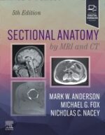
Sectional Anatomy by MRI and CT – Anderson
$1,495,000A sure grasp of cross-sectional anatomy is essential for accurate radiologic interpretation, and Sectional Anatomy by MRI and CT, 5th Edition, provides exactly the information needed in a highly illustrated, quick-reference format. New coverage of the cervical spine, brain, and thumb, as well as new on/off labels in the eBook version make this title an essential diagnostic tool for both residents and practicing radiologists.
Key Features
Features color-coded labels for nerves, vessels, muscles, bone tendons, and ligaments that facilitate accurate identification of key anatomic structures.
Provides new on/off labels in the accompanying eBook, as well as scroll and zoom capabilities on photos for convenient access during interpretation sessions and real-time resident education.
Presents carefully labeled MRIs for all body parts, as well as schematic diagrams and concise statements, to clarify correlations between bones and tissues.
Includes CT scans for selected body parts to enhance anatomic visualization.
Features 1165 state-of-the-art images that can be viewed in three standard planes: axial, coronal, and sagittal.
An eBook version is included with purchase. The eBook allows you to access all of the text, figures, and references, with the ability to search, customize your content, make notes and highlights, and have content read aloud. Any additional digital ancillary content may publish up to 6 weeks following the publication date. -
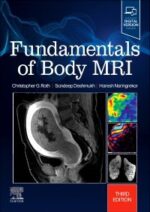
Fundamentals of Body MRI, 3rd Edition – Roth
$485,000Effectively perform and interpret MR body imaging with this concise, highly illustrated resource! Fundamentals of Body MRI, 3rd Edition, covers the essential concepts residents, fellows, and practitioners need to know, laying a solid foundation for understanding the basics and making accurate diagnoses. This easy-to-use title in the Fundamentals of Radiology series covers all common body MR imaging indications and conditions, while providing new content on body MRI emergencies, physics, and noninterpretive skills with an emphasis on quality and safety.
Key Features
Covers all common body MR imaging content, along with discussion of how physics, techniques, hardware, and artifacts affect results—all summarized in an easy-to-read format with practical applications throughout.Features more than 1,600 detailed MRI images and 100 algorithms and diagrams that highlight key findings and help you grasp visual nuances of images you’re likely to encounter.
Contains extensively revised content on liver lesions, including new coverage on LI-RADS system, and new safety tips and guidelines that keep you up to date.
Includes new information on MR defecography and advances in rectal cancer staging and post-treatment imaging, including new content on inflammatory bowel disease.
An eBook version is included with purchase. The eBook allows you to access all of the text, figures and references, with the ability to search, customize your content, make notes and highlights, and have content read aloud. Any additional digital ancillary content may publish up to 6 weeks following the publication date.
-
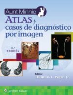
Aunt Minnie. Atlas y casos de diagnóstico por imagen – Pope
$517,000Características principales:
• Cubre todas las subespecialidades radiológicas e incluye imágenes, historia, hallazgos, diagnóstico y discusión
• Proporciona «Perlas» al final de cada caso que ayudan a reforzar las características clave y ofrecen una revisión rápida de los principales puntos destacados
• Presenta muchos casos clásicos nuevos con imágenes actualizadas e imágenes «avanzadas» adicionales en varios casos
• Escrito por subespecialistas de renombre y editado por el mundialmente conocido Dr. Thomas L. Pope$609,000 -

Advanced Imaging in Ischemic and Hemorrhagic Stroke – Gemmete
$455,000In this issue of Neuroimaging Clinics, guest editors Drs. Joseph Gemmete and Zachary Wilseck bring their considerable expertise to the topic of Advanced Imaging in Ischemic and Hemorrhagic Stroke. Top experts in the field discuss metabolic imaging such as PET, in vivo O17 MRS imaging, quantitative sodium MR imaging, pharmacological MRI (pHMRI), dual-energy CT, vessel wall imaging, and more.Key Features
- Contains 11 relevant, practice-oriented topics including CT/CTA/perfusion in acute ischemic stroke/vasospasm; MRI/MRA/perfusion in acute ischemic stroke and vasospasm; MR imaging of cerebral aneurysmal wall for rupture risk; dual-energy CT in imaging of acute ischemic and hemorrhagic stroke; metabolic imaging of acute ischemic stroke (PET, spectroscopy, O17 imaging, sodium imaging, PH imaging); and more.
- Provides in-depth clinical reviews on advanced imaging in ischemic and hemorrhagic stroke, offering actionable insights for clinical practice.
- Presents the latest information on this timely, focused topic under the leadership of experienced editors in the field. Authors synthesize and distill the latest research and practice guidelines to create clinically significant, topic-based reviews.
-
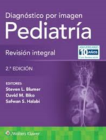
Diagnóstico por imagen. Pediatría: Revisión integral – Blumer
$375,000Diagnóstico por imagen. Pediatría: Revisión integral, del Dr. Steven L. Blumer, es una herramienta de revisión práctica y actualizada que abarca el amplio campo de la radiología pediátrica, escrita específicamente para el examen de certificación de la subespecialidad. Para una buena preparación de los estudiantes, este útil recurso contiene más de 300 preguntas de opción múltiple, ricas en imágenes, con explicaciones detalladas de las respuestas correctas e incorrectas, contenido totalmente revisado y tablas de alto rendimiento para una fácil revisión.
Esta 2.ª edición se basa en el éxito de la primera, y cubre todos los aspectos esenciales de la imagenología pediátrica de manera que sirva como guía para residentes, quienes podrán evaluar su conocimiento y revisar el material en un formato similar al examen central de la American Board of Radiology (ABR) o los exámenes de consejos o instituciones similares locales. La obra cubre ocho áreas esenciales de la radiología pediátrica: tubo digestivo, aparato genitourinario, sistema musculoesquelético, neurorradiología pediátrica, y radiología de tórax, vascular, cardíaca y de múltiples sistemas.$440,000 -
Ecografía Diagnóstica y Anatómica MSK – Jimenez
$278,000Pieza básica y esencial para el desarrollo en el conocimiento de la Ecografía Musculoesquelética (MSK) para todos aquellos que deseen iniciarse o estén iniciándose en esta especialidad tan de moda. Dirigido tanto para traumatólogos como para técnicos radiólogos, fisioterapeutas, rehabilitadores y todo aquel que se quiera relacionar directamente con el aparato locomotor y familiarizarse con la «Eco MSK» para el diagnóstico y tratamiento de los procesos traumatológicos. Dividido en seis secciones por articulaciones, ECOGRAFÍA Diagnóstica y Anatómica MSK tiene una estructura bien definida con un completo recorrido por la anatomía ecográfica y las técnicas de examen para el diagnóstico clínico, con multitud de imágenes figurativas y de exploraciones a todo color para entender rápidamente la correlación entre la posición del transductor y la imagen ecográfica. Sin duda, la herramienta fundamental que va a ayudar a los menos expertos a realizar una correcta exploración y comprender la utilidad de la ecografía, iniciando el camino en el gran universo Eco MSK del Profesor JF Jiménez Díaz.
-
Radiología de Mama – Joe
$357,000Centrándose en la información práctica, Radiología de mama, 4.ª edición, hace hincapié en los fundamentos básicos para ayudarle a comprender los conceptos principales de las imágenes de mama, refrescar sus conocimientos sobre conceptos clave y aprender estrategias para proporcionar informes provechosos a los médicos solicitantes. Esta edición, completamente reescrita y reorganizada, enfatiza los conocimientos esenciales necesarios en un formato de fácil lectura, con actualizaciones exhaustivas que cubren las nuevas modalidades de imagen, las últimas directrices y la integración de la información relativa a los conceptos físicos a lo largo de todo el libro.
$420,000 -
Kandarpa. Manual de procedimientos en radiología intervencionista 6ed. 2024
$398,000Kandarpa. Manual de procedimientos en radiología intervencionista es una referencia indispensable para médicos intervencionistas de todos los niveles de experiencia. Desde su primera edición en 1989, ha seguido el rápido crecimiento y cambio de la disciplina, con más de 100 capítulos que reflejan la expansión y avance del campo. Además de detallar procedimientos comunes, resalta los roles emergentes de los tratamientos endovasculares y no vasculares para diversas enfermedades. Es ideal no solo para la práctica de radiólogos intervencionistas y generales, sino también para médicos residentes en formación, profesionales de enfermería y técnicos en procedimientos especiales.
En esta 6.ª edición se han ampliado los capítulos clave sobre calidad, manejo de riesgos y seguridad, así como control de radiación e infecciones, lo que refleja la evolución del campo. También se enfoca en el manejo completo del paciente antes, durante y después del procedimiento, lo que brinda información esencial en un formato fácil de usar. Cada procedimiento incluye indicaciones, contraindicaciones, preparación, técnica, manejo posterior y prevención de complicaciones.


